. 2011:2011:325789.
doi: 10.1155/2011/325789. Epub 2011 Jun 28.

Ultrastructural alterations of myelinated fibers and oligodendrocytes in the prefrontal cortex in schizophrenia: a postmortem morphometric study

Affiliations

Ultrastructural alterations of myelinated fibers and oligodendrocytes in the prefrontal cortex in schizophrenia: a postmortem morphometric study

Natalya A Uranova et al. Schizophr Res Treatment. 2011.

Abstract

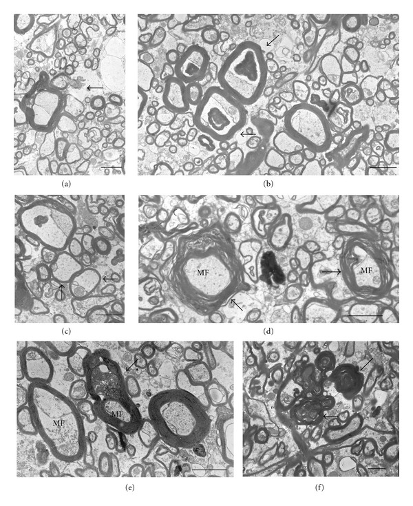
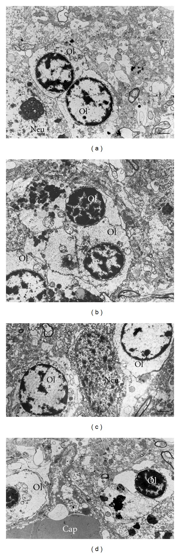
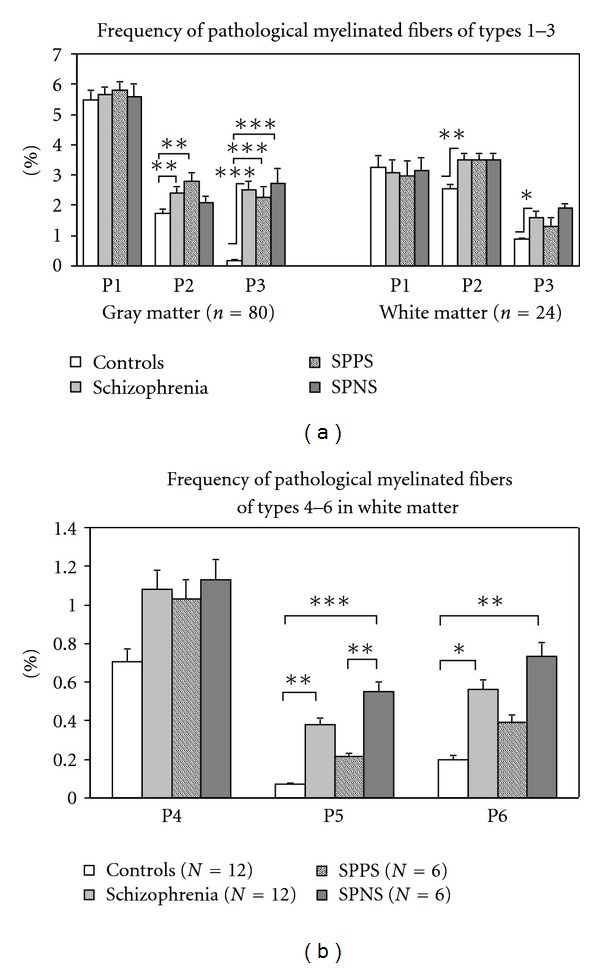
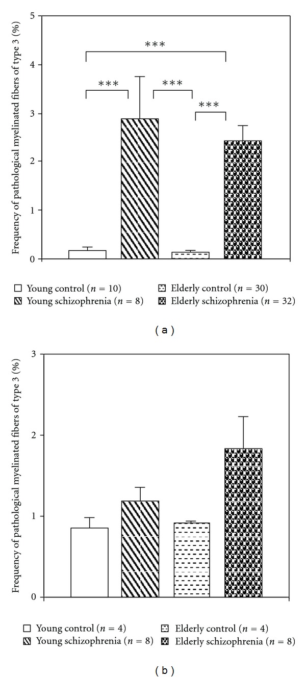
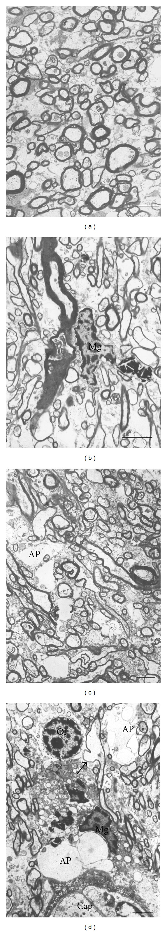
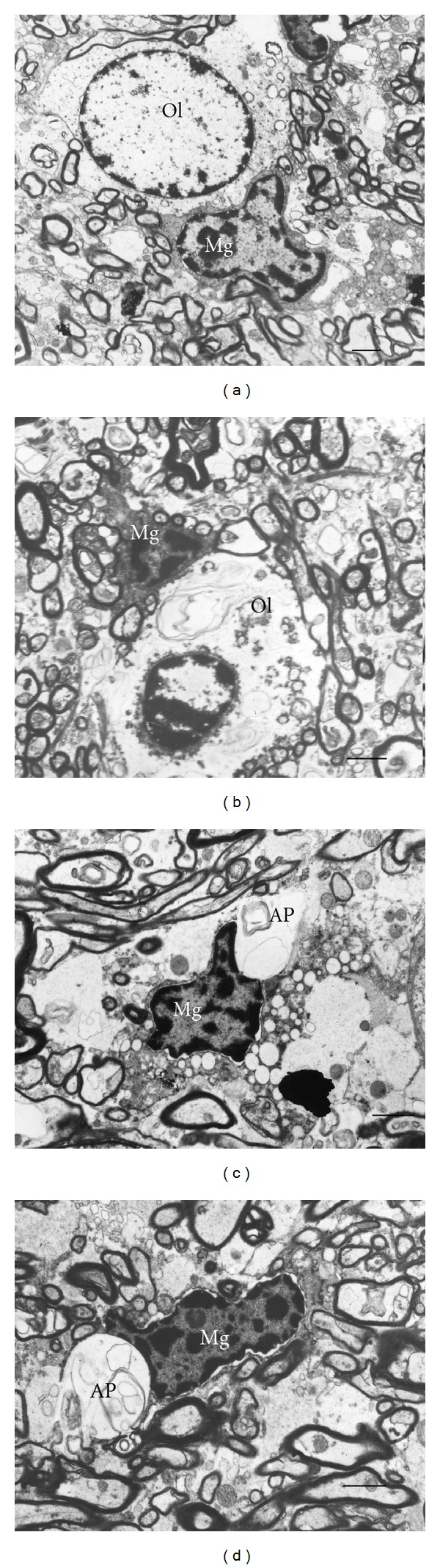
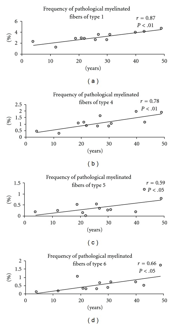
Schizophrenia is believed to result from altered neuronal connectivity and impaired myelination. However, there are few direct evidence for myelin abnormalities in schizophrenia. We performed electron microscopic study of myelinated fibers and oligodendrocytes and morphometric study of myelinated fibers in the prefrontal cortex in gray and white matters in schizophrenia and normal controls. Six types of abnormal fibers and ultrastructural alterations of oligodendrocytes were found in schizophrenia. No significant group differences in area density of myelinated fibers were found. Frequency of pathological fibers was increased significantly in gray matter in young and elderly schizophrenia patients and in patients with predominantly positive symptoms. In contrast, in white matter, frequency of altered fibers was increased significantly in elderly patients, in patients with predominantly negative symptoms, and correlated with illness duration. Progressive alterations of myelinated fibers in white matter might be followed by alterations of myelinated fibers in gray matter in schizophrenia.


Figures

Figure 1
Figure 1
Six types of pathological myelinated fibers. Descriptions in the text. Scale bars  =  2 μm.
Figure 2
Figure 2
Electron micrographs of oligodendrocytes in gray matter from control subject (a) and from subjects with schizophrenia (b–d). Dystrophic alterations of oligodendrocytes, Ol, (b, c) and destructive changes of oligodendrocytes (d). Scale bars  = 2 μm.
Figure 3
Figure 3
Frequency of pathological myelinated fibers present in gray matter and in white matter (mean ± SEM). SPPS: schizophrenia subjects with predominantly positive symptoms. SPNS: schizophrenia subjects with predominantly negative symptoms. *P < .05, **P < .01, ***P < .001.
Figure 4
Figure 4
Histograms demonstrating the results of comparisons of the frequency of pathological myelinated fibers of type 3 in young and elderly patient subgroups with young and elderly controls. (a) Gray matter, (b) White matter (mean ± SEM).
Figure 5
Figure 5
Electron micrographs of white matter from control brain (a) and from the brains of subjects with schizophrenia (b–d). Both thinly myelinated (b, c) and axons with focal lysis of myelin sheath (d) as well as some axons with abnormally thick myelin sheaths (Figure 1(b)) were observed in schizophrenia in contrast to controls. Ap: astrocytic process, Mg: microglial cell. Ol: oligodendrocyte. Scale bars = 2 μm.
Figure 6
Figure 6
Electron micrographs of oligodendrocytes and microglia in white matter from control subjects (a) and from subjects with schizophrenia (b–d). Dystrophic changes of oligodendrocyte, Ol, (b). Activated microglia in close apposition to swollen astrocytic process (Ap) containing myelin debris in schizophrenia brain (c) Microglial cell participates in phagocytosis of myelin membranous debris in schizophrenia (d). Scale bars = 2 μm.
Figure 7
Figure 7
The association between frequency of pathological myelinated fibers in white matter and duration of schizophrenia.

References

    1. Davis KL, Stewart DG, Friedman JI, et al. White matter changes in schizophrenia evidence for myelin-related dysfunction. Archives of General Psychiatry. 2003;60(5):443–456. - PubMed
    1. Bartzokis G, Nuechterlein KH, Lu PH, Gitlin M, Rogers S, Mintz J. Dysregulated brain development in adult men with schizophrenia: a magnetic resonance imaging study. Biological Psychiatry. 2003;53(5):412–421. - PubMed
    1. Bartzokis G, Lu PH, Stewart SB, et al. In vivo evidence of differential impact of typical and atypical antipsychotics on intracortical myelin in adults with schizophrenia. Schizophrenia Research. 2009;113(2-3):322–331. - PMC - PubMed
    1. Kubicki M, McCarley RW, Shenton ME. Evidence for white matter abnormalities in schizophrenia. Current Opinion in Psychiatry. 2005;18(2):121–134. - PMC - PubMed
    1. Kubicki M, McCarley R, Westin CF, et al. A review of diffusion tensor imaging studies in schizophrenia. Journal of Psychiatric Research. 2007;41(1-2):15–30. - PMC - PubMed

LinkOut - more resources