

A rare form of melanoma masquerading as a diabetic foot ulcer: a case report
- PMID: 22937296
- PMCID: PMC3420798
- DOI: 10.1155/2012/502806
A rare form of melanoma masquerading as a diabetic foot ulcer: a case report
Abstract
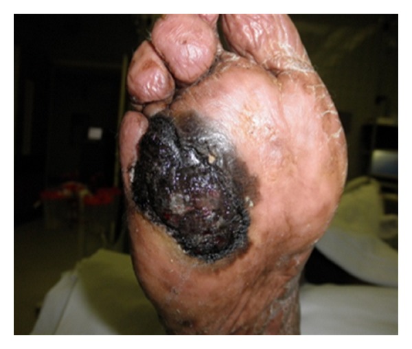
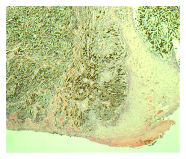
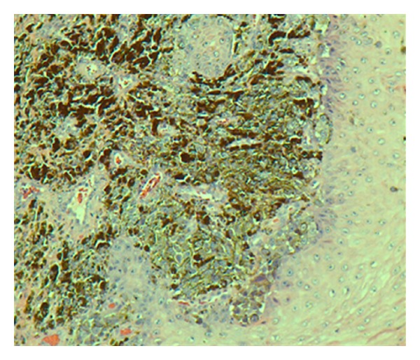
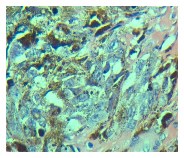
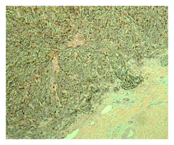
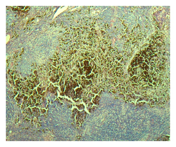
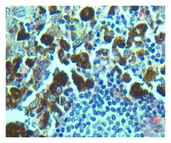
Background. Acral lentiginous melanoma (ALM) is a less-common form of melanoma in US, and it accounts for about 5% of all diagnosed melanomas in US. ALM is often overlooked until it is well advanced because of the lesion's location and its atypical appearance in the early stages. We present a case of ALM initially presented as a diabetic foot ulcer. Case Report. An 81-year-old man initially presented to the primary care clinic with a right foot diabetic ulcer. There was a large plantar, dark-colored ulcer that bled easy. Initial excision biopsy revealed Clark's Level IV ALM. Subsequent definitive wide excision and sentinel node biopsy confirmed ALM with metastasis to inguinal lymph nodes (stage IIIb). The treatment included wide margin excision of the lesion with en bloc amputations of 4th and 5th toes, followed by adjuvant chemotherapy. Discussion. The development of ALM may potentially relate to diabetes as a reported higher prevalence of diabetes with ALM patients. Conclusion. The difficulty in early diagnosing of ALM remains as a formidable challenge particularly in diabetic patients who commonly develop plantar foot ulcers due to the diabetic neuropathy. This case reiterates the importance of a thorough foot exam in such patients.
Figures
Similar articles
-
Progression from Acral Lentiginous Melanoma in situ to Invasive Acral Lentiginous Melanoma.Ann Dermatol. 2009 May;21(2):185-8. doi: 10.5021/ad.2009.21.2.185. Epub 2009 May 31. Ann Dermatol. 2009. PMID: 20523783 Free PMC article.
-
Acral lentiginous melanoma versus other melanoma: A single-center analysis in Japan.J Dermatol. 2017 Aug;44(8):932-938. doi: 10.1111/1346-8138.13834. Epub 2017 Mar 24. J Dermatol. 2017. PMID: 28342269
-
Malignant melanoma misdiagnosed as diabetic foot ulcer: A case report.Medicine (Baltimore). 2017 Jul;96(29):e7541. doi: 10.1097/MD.0000000000007541. Medicine (Baltimore). 2017. PMID: 28723771 Free PMC article.
-
Diagnosis and Management of Acral Lentiginous Melanoma.Curr Treat Options Oncol. 2018 Jun 27;19(8):42. doi: 10.1007/s11864-018-0560-y. Curr Treat Options Oncol. 2018. PMID: 29951919 Review.
-
Metastatic acral lentiginous melanoma in a tertiary referral center in Switzerland: a systematic analysis.Melanoma Res. 2018 Oct;28(5):442-450. doi: 10.1097/CMR.0000000000000465. Melanoma Res. 2018. PMID: 29847461
Cited by
-
Acral melanoma with satellitosis, disguised as a longstanding diabetic ulcer: a great mimicry.Int Wound J. 2016 Oct;13(5):1006-8. doi: 10.1111/iwj.12481. Epub 2015 Sep 24. Int Wound J. 2016. PMID: 26400657 Free PMC article.
-
Misdiagnosis of diabetic foot ulcer in patients with undiagnosed skin malignancies.Int Wound J. 2022 May;19(4):871-887. doi: 10.1111/iwj.13688. Epub 2021 Oct 29. Int Wound J. 2022. PMID: 34713964 Free PMC article.
References
-
- Reed R. Acral lentiginous melanoma. In: Hartmann W, Reed R, editors. New Concepts in Surgical Pathology of the Skin. New York, NY, USA: Wiley; 1976. pp. 89–90.
-
- Hudson DA, Krige JEJ, Stubbings H. Plantar melanoma: results of treatment in three population groups. Surgery. 1998;124(5):877–882. - PubMed
-
- Chen YJ, Wu CY, Chen JT, Shen JL, Chen CC, Wang HC. Clinicopathologic analysis of malignant melanoma in Taiwan. Journal of the American Academy of Dermatology. 1999;41(6):945–949. - PubMed
-
- Luk NM, Ho LC, Choi CL, Wong KH, Yu KH, Yeung WK. Clinicopathological features and prognostic factors of cutaneous melanoma among Hong Kong Chinese. Clinical and Experimental Dermatology. 2004;29(6):600–604. - PubMed
-
- Ishihara K, Saida T, Yamamoto A. Updated statistical data for malignant melanoma in Japan. International Journal of Clinical Oncology. 2001;6(3):109–116. - PubMed
Publication types
Grants and funding
LinkOut - more resources
Full Text Sources
Research Materials
