Case Reports
. 2011:2011:176546.
doi: 10.1155/2011/176546. Epub 2011 Jul 14.

Inflammatory pseudotumor of the head presenting with hemiparesis and aphasia

Affiliations
Case Reports

Inflammatory pseudotumor of the head presenting with hemiparesis and aphasia

K Saifudheen et al. Case Rep Neurol Med. 2011.

Abstract

Inflammatory pseudotumor most commonly occurs in the orbit and produces orbital pseudotumor, but extension into brain parenchyma is uncommon. We report a case of inflammatory pseudotumor involving sphenoid sinus, cavernous sinus, superior orbital fissure, orbital muscle, and intracranial extension into left temporal lobe producing right hemiparesis and wernicke's aphasia. The patient improved clinically and radiologically with steroid administration. This paper provides an insight into the spectrum of involvement of inflammatory pseudotumor and the importance of early diagnosis of the benign condition.


Figures
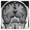
Figure 1
Figure 1
Coronal T1-weighted contrast image shows enhancing lesion in the cavernous sinus bilaterally with sphenoid sinus fullness (black arrow).
Figure 2
Figure 2
Coronal T1-weighted contrast image shows proptosis of the right globe. There is intense enhancement and enlargement of the right superior and lateral recti (black arrow).
Figure 3
Figure 3
Coronal and Axial T1-weighted contrast image shows intensely enhancing extra-axial mass lesion extending from left cavernous area compressing and invading left anterior temporal lobe (black arrow). There is significant perilesional edema. Lateral ventricle is compressed with midline shift to the right.
Figure 4
Figure 4
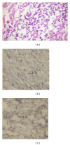
(a) Histopathology showing mature lymphocyte infiltration with occasional macrophage; (b) and (c) immunohistochemical staining for CD3 and CD20 showing the CD3 and CD20 positive lymphocyte.
Figure 5
Figure 5
Axial T1-weighted contrast image shows almost complete resolution of the temporal lobe lesion with mild dilatation of the left temporal horn.

References

    1. Lee JH, Kim K, Chung SW, et al. A case report of inflammatory peudotumor involving the clivus: CT and MR findings. Korean Journal of Radiology. 2001;2(4):231–234. - PMC - PubMed
    1. De Vuysere S, Hermans R, Sciot R, et al. Extraorbital inflammatory pseudotumor of the head and neck: CT and MR findings in three patients. American Journal of Neuroradiology. 1999;20(6):1133–1139. - PMC - PubMed
    1. Narla LD, Newman B, Spottswood SS, et al. Inflammatory pseudotumor. Radiographics. 2003;23(3):719–729. - PubMed
    1. Lee EJ, Jung SL, Kim BS, et al. MR imaging of orbital inflammatory pseudotumors with extraorbital extension. Korean Journal of Radiology. 2005;6(2):82–88. - PMC - PubMed
    1. Bramwit M, Kalina P, Rustia-Villa M. Inflammatory pseudotumor of the choroid plexus. American Journal of Neuroradiology. 1997;18(7):1307–1309. - PMC - PubMed

Publication types

LinkOut - more resources