

Extra-abdominal fibromatosis (desmoid tumor): a rare tumor of the lower extremity arising from the popliteal fossa
- PMID: 22937461
- PMCID: PMC3420745
- DOI: 10.1155/2011/184906
Extra-abdominal fibromatosis (desmoid tumor): a rare tumor of the lower extremity arising from the popliteal fossa
Abstract
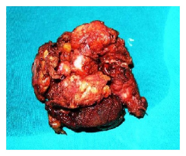
Aggressive fibromatosis is a rare soft tissue tumor. Although it lacks metastatic potential, it can grow aggressively in a locally infiltrating pattern. The tumors frequently recur after surgical excision, which remains the treatment of choice. Optional combinations of radiotherapy and/or chemotherapy have been used postoperatively for recurrent disease and/or inoperable cases. A palpable mass was detected in the popliteal fossa of the right lower extremity in a 48-year-old man. Magnetic resonance imaging showed a contrast-enhancing noncalcified lesion initially felt to represent a vascular tumor. An invasive mass adherent to the surrounding tissue was visualized intraoperatively and extensively debulked. The patient's postoperative course was uneventful. Histologic examination of the surgical specimen was consistent with an extra-abdominal desmoid tumor. After appropriate recognition, wide local excision may be the most appropriate treatment for fibromatosis of the extremity. However, the rarity of this tumor and the difficulty inherent in distinguishing it from similar-appearing tumors are necessitating histologic confirmation of the diagnosis.
Figures


Similar articles
-
A Rare Case of Extra-abdominal Desmoid-type Fibromatosis Arising from the Popliteal Fossa.Cureus. 2018 Nov 28;10(11):e3651. doi: 10.7759/cureus.3651. Cureus. 2018. PMID: 30723650 Free PMC article.
-
Fibromatosis with aggressive demeanor: Benign impersonator of malignancy.World J Nucl Med. 2020 Dec 12;20(1):121-124. doi: 10.4103/wjnm.WJNM_55_20. eCollection 2021 Jan-Mar. World J Nucl Med. 2020. PMID: 33850503 Free PMC article.
-
Extra-abdominal fibromatosis (desmoid tumor) arising in the infratemporal fossa: a case report.Skull Base Surg. 1998;8(4):237-41. doi: 10.1055/s-2008-1058191. Skull Base Surg. 1998. PMID: 17171074 Free PMC article.
-
An unusual finding in a desmoid-type fibromatosis of the pancreas: a case report and review of the literature.J Med Case Rep. 2018 May 12;12(1):123. doi: 10.1186/s13256-018-1635-x. J Med Case Rep. 2018. PMID: 29751773 Free PMC article. Review.
-
A rare presentation of a large extra-abdominal desmoid tumor of the posterior neck and back.Am J Otolaryngol. 2013 Nov-Dec;34(6):727-30. doi: 10.1016/j.amjoto.2013.08.013. Epub 2013 Sep 13. Am J Otolaryngol. 2013. PMID: 24035615 Review.
Cited by
-
Desmoid Fibromatosis of the Triceps and Sternocleidomastoid-A Report of Two Cases.J Orthop Case Rep. 2025 Jul;15(7):201-205. doi: 10.13107/jocr.2025.v15.i07.5824. J Orthop Case Rep. 2025. PMID: 40635923 Free PMC article.
-
A Rare Case of Extra-abdominal Desmoid-type Fibromatosis Arising from the Popliteal Fossa.Cureus. 2018 Nov 28;10(11):e3651. doi: 10.7759/cureus.3651. Cureus. 2018. PMID: 30723650 Free PMC article.
-
Largest size of extra-abdominal fibromatosis of axilla in a young man.BMJ Case Rep. 2019 Aug 1;12(8):e230670. doi: 10.1136/bcr-2019-230670. BMJ Case Rep. 2019. PMID: 31375508 Free PMC article. No abstract available.
References
-
- MacFarlane J. Clinical Reports of Surgical Practice of Glasgow Royal Infirmary. Glasgow, Scotland: 1832.
-
- Brenner P, Rammelt S. Abdominal wall and foot reconstruction after extensive desmoid tumor resection with free tissue transfer. Langenbeck’s Archives of Surgery. 2002;386(8):592–597. - PubMed
-
- Barbashina V, Karabakhtsian R, Aisner S, Bolanowski P, Patterson F, Hameed M. Desmoplastic fibroma of the rib. Archives of Pathology and Laboratory Medicine. 2002;126(6):721–722. - PubMed
-
- Rosen PP, Oberman HA. Atlas of Tumor Pathology. Tumors of the Mammary Gland. Washington, DC, USA: AFIP; 1993.
-
- Winer-Muram HT, Bowman LC, Parham D. Intrathoracic desmoid tumor: CT and MRI appearance. Southern Medical Journal. 1994;87(10):1007–1009. - PubMed
Publication types
LinkOut - more resources
Full Text Sources
