Review
. 2012 Dec;103(12):2039-44.
doi: 10.1111/cas.12007. Epub 2012 Oct 10.

Contribution of GATA1 dysfunction to multi-step leukemogenesis

Affiliations
Review

Contribution of GATA1 dysfunction to multi-step leukemogenesis

Ritsuko Shimizu et al. Cancer Sci. 2012 Dec.

Abstract

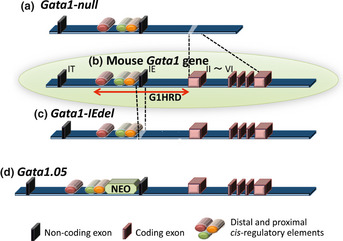
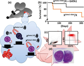
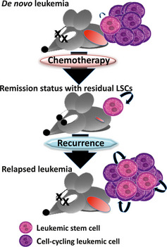
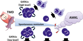
In mammals, hematopoietic homeostasis is maintained by a fine-tuned balance among the self-renewal, proliferation, differentiation and survival of hematopoietic stem cells and their progenies. Each process is also supported by the delicate balance of the expression of multiple genes specific to each process. GATA1 is a transcription factor that comprehensively regulates the genes that are important for the development of erythroid and megakaryocytic cells. Accumulating evidence supports the notion that defects in GATA1 function are intimately linked to hematopoietic disorders. In particular, the somatic mutation of the GATA1 gene, which leads to the production of N-terminally truncated GATA1, contributes to the genesis of transient myeloproliferative disorder and acute megakaryoblastic leukemia in infants with Down syndrome. Similarly, a mutation in the GATA1 regulatory region that reduces GATA1 expression is involved in the onset of erythroid leukemia in mice. In both cases, the accumulation of immature progenitor cells caused by GATA1 dysregulation underlies the pathogenesis of the leukemia. This review provides a summary of multi-step leukemogenesis with a focus on GATA1 dysfunction.


Figures

Figure 1
Figure 1
The structure of the mouse Gata1 gene. The mouse Gata1 gene is composed of two alternative non‐coding first exons and five constitutive coding exons (b; inside the green circle). G1HRD is composed of three cis‐regulatory regions (shown by red, green and orange bars) located upstream of the IE exon and the first intron. The Gata1‐null (a) and Gata1‐IEdel (b) alleles were constructed by deleting the five coding exons or the IE exon, respectively. The insertion of a Neo cassette between the cis‐regulatory elements and the IE exon reduces the gene expression of the Gata1.05 allele (d).
Figure 2
Figure 2
Multi‐step leukemogenesis caused by the dysregulation of Gata1 gene expression. (a) There are two types of erythroid progenitor cells in females heterozygous for the Gata1 gene mutation due to the random inactivation of the X chromosome. Erythroid progenitor cells with the activated wild‐type allele undergo terminal maturation, whereas those with the inactivated wild‐type allele fail to differentiate and continue to proliferate. Immature cells with the activated Gata1‐null allele die by apoptosis due to the absence of GATA1. In contrast, a low level of GATA1 expression elongates the lifespan of the progenitor cells expressing either the activated Gata1‐IEdel or Gata1.05 allele. Consequently, those cells may acquire additional genetic event(s). (b) Survival curves of Gata1‐IEdel heterozygous females with (black line; = 35) or without (red line; = 10) the G1HRDGATA1 transgene. (c) A typical flow cytometry dot plot showing leukemic cell population positive for c‐Kit and CD71 (right panel). Left panel depicts a graph for side‐scatter (SSC) versus forward‐scatter (FSC). Leukemic cells in peripheral blood are gated in a red circle. (d) Leukemic cells in bone marrow of Gata1‐IEdel heterozygous females are morphologically similar to proerythroblasts.
Figure 3
Figure 3
Pernicious changes in leukemic stem cells (LSCs) in relapsed leukemia. Most LSCs are quiescent, and the number of LSCs is maintained at a steady state in a de novo leukemia. After eliminating proliferating leukemic cells with inappropriate chemotherapy, the residual LSCs are stimulated to enter the cell cycle and produce siblings and progenies (remission status with residual LSCs). Upon relapsing to full‐blown leukemia, the LSCs fail to return to quiescence and are maintained at a higher number than before (relapsed leukemia).
Figure 4
Figure 4
Multi‐step leukemogenesis caused by a structural mutation in the GATA1 protein. Because the contribution of the GATA1‐S mutation to the hyper‐proliferative phenotype of megakaryocytes is restricted to fetal hematopoiesis, the phenotypes observed in babies with Down syndrome with transient myeloproliferative disorder (DSTMD) and mice rescued with GATA1‐S disappear spontaneously. At high levels of expression, GATA1‐S has the potential to stimulate megakaryocytic differentiation and rapidly eliminate accumulated abnormal megakaryocytic progenitor cells. However, a low level of GATA1‐S fails to promote differentiation and instead extends the life spans of those progenitor cells bearing the GATA1‐S mutation. Consequently, those cells have an opportunity to acquire additional genetic hit(s) and eventually transform into a leukemic cell.

Similar articles

Cited by

References

    1. Tsai SF, Martin DI, Zon LI, D'Andrea AD, Wong GG, Orkin SH. Cloning of cDNA for the major DNA‐binding protein of the erythroid lineage through expression in mammalian cells. Nature 1989; 339: 446–51. - PubMed
    1. Yamamoto M, Ko LJ, Leonard MW, Beug H, Orkin SH, Engel JD. Activity and tissue‐specific expression of the transcription factor NF‐E1 multigene family. Genes Dev 1990; 4: 1650–62. - PubMed
    1. Shimizu R, Yamamoto M. Gene expression regulation and domain function of hematopoietic GATA factors. Semin Cell Dev Biol 2005; 16: 129–36. - PubMed
    1. Zheng W, Flavell RA. The transcription factor GATA‐3 is necessary and sufficient for Th2 cytokine gene expression in CD4 T cells. Cell 1997; 89: 587–96. - PubMed
    1. Suzuki M, Shimizu R, Yamamoto M. Transcriptional regulation by GATA1 and GATA2 during erythropoiesis. Int J Hematol 2011; 93: 150–5. - PubMed

Publication types

MeSH terms

Substances