Review
. 2013 Dec 1;19(16):1940-52.
doi: 10.1089/ars.2012.4847. Epub 2012 Oct 15.

Redox and reactive oxygen species regulation of mitochondrial cytochrome C oxidase biogenesis

Affiliations
Review

Redox and reactive oxygen species regulation of mitochondrial cytochrome C oxidase biogenesis

Myriam Bourens et al. Antioxid Redox Signal. .

Abstract

Significance: Cytochrome c oxidase (COX), the last enzyme of the mitochondrial respiratory chain, is the major oxygen consumer enzyme in the cell. COX biogenesis involves several redox-regulated steps. The process is highly regulated to prevent the formation of pro-oxidant intermediates.

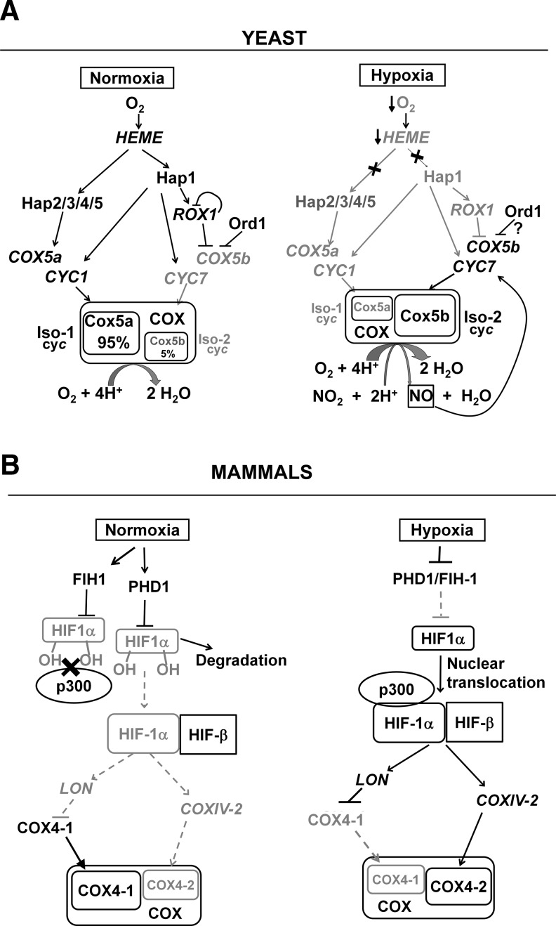
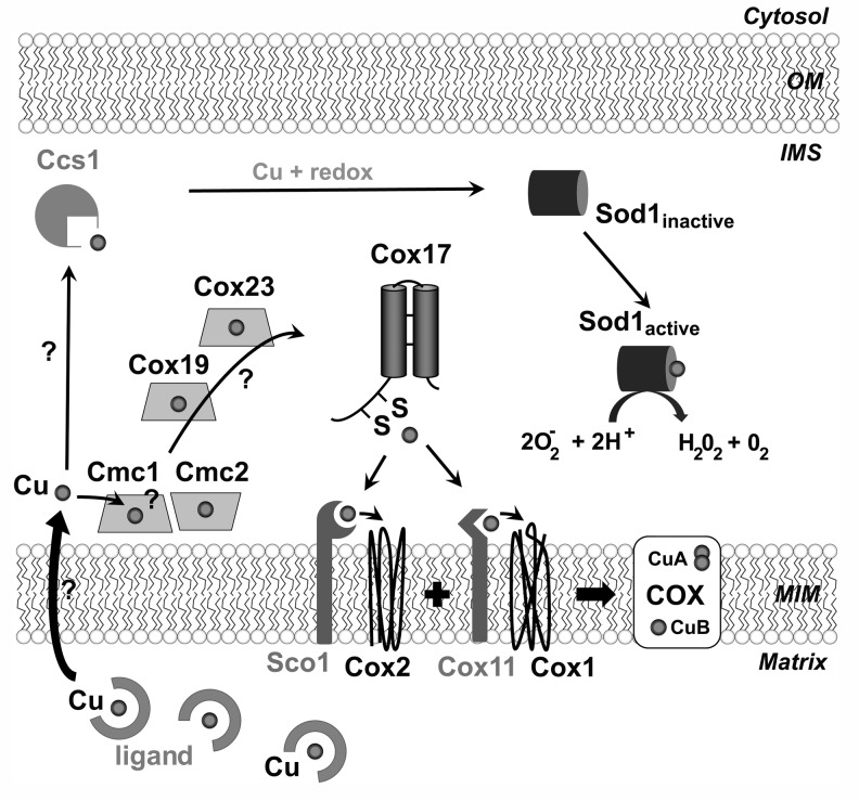
Recent advances: Regulation of COX assembly involves several reactive oxygen species and redox-regulated steps. These include: (i) Intricate redox-controlled machineries coordinate the expression of COX isoenzymes depending on the environmental oxygen concentration. (ii) COX is a heme A-copper metalloenzyme. COX copper metallation involves the copper chaperone Cox17 and several other recently described cysteine-rich proteins, which are oxidatively folded in the mitochondrial intermembrane space. Copper transfer to COX subunits 1 and 2 requires concomitant transfer of redox power. (iii) To avoid the accumulation of reactive assembly intermediates, COX is regulated at the translational level to minimize synthesis of the heme A-containing Cox1 subunit when assembly is impaired.

Critical issues: An increasing number of regulatory pathways converge to facilitate efficient COX assembly, thus preventing oxidative stress.

Future directions: Here we will review on the redox-regulated COX biogenesis steps and will discuss their physiological relevance. Forthcoming insights into the precise regulation of mitochondrial COX biogenesis in normal and stress conditions will likely open future perspectives for understanding mitochondrial redox regulation and prevention of oxidative stress.


Figures

FIG. 1.
FIG. 1.
Cytochrome c oxidase (COX): metal cofactors and function. (A) Metal cofactors in COX catalytic subunits 1 and 2. The catalytic core of COX contains copper and iron prosthetic groups. Two copper atoms bound to subunit 2 constitute the CuA site. In subunit 1, there are two redox centers: a low-spin heme a group, and a binuclear center formed by a third copper atom, CuB, associated with the high-spin heme a3 group. The two heme planes are both essentially perpendicular to the mitochondrial membrane plane (not depicted here, but see panel B). Ligand-bound structures of bovine COX subunits 1 and 2 were obtained from the protein data bank (PDB) website (www.rcsb.org/pdb/home/home.do) and modified to prepare the figure. The YASARA molecular-graphics, -modeling, and -simulation program, developed by Elmer Krieger, was used to generate a cartoon model of the different proteins. (B) COX functions as an electron-driven proton pump. The CuA site in subunit 2 is the primary acceptor of electrons from ferrocytochrome c. Electrons are subsequently transferred to the low-spin heme a group located in subunit 1. And from there to the binuclear CuB-heme a3 center of subunit 1, where oxygen binds and is sequentially reduced to water. Electron transfer to dioxygen is coupled to proton pumping across the inner mitochondrial membrane that contributes generating a gradient that is used by the F1F0-ATPase to synthesize adenosine-5′-triphosphate (ATP). In addition, one substrate proton per electron (not depicted here) is delivered into the binuclear site to form water.
FIG. 2.
FIG. 2.
Reactive oxygen species (ROS) and redox control mechanisms involved in COX biogenesis. General chaperones and RNA-specific translational activators (not depicted here) are required for synthesis of the mtDNA-encoded subunits forming the COX catalytic core. Following their insertion into the inner membrane (MIM), Cox1 and Cox2 are matured by addition of metal cofactors. At some point, substrate-specific chaperones bind Cox1, Cox2, and Cox3 to maintain them in an assembly-competent state. Following Cox1 maturation, the nuclear DNA-encoded Cox5 and 6 subunits are added to Cox1 before incorporation of the other core subunits and the rest of the accessory subunits to form the holoenzyme. The ROS and redox control mechanisms involved in COX biogenesis are noted in start-shape boxes.
FIG. 3.
FIG. 3.
Mechanisms of oxygen-regulated COX subunit isoform switch in yeast and mammals. (A) Transcriptional regulation of COX5a/COX5b in the yeast Saccharomyces cerevisiae. Normoxic (>0.5 μM O2) repression of COX5b is mediated through a heme-dependent pathway. In the presence of oxygen, heme is synthesized and binds to the transcriptional factor Hap1 leading to its activation. Hap1 induces the expression of a set of OXPHOS genes, including CYC1 and CYC7. It also activates the expression of the general transcription repressor Rox1, which binds to a consensus ATTGTTCTC box on the promoter region of anaerobic genes, such as COX5b, CYC7, and itself to repress their expression. Hap1 acts on CYC7 and ROX1 as an activator directly and also as a repressor through Rox1. This feedback regulatory loop has been proposed to confer mutational robustness in yeast transcription factor regulation (25). Additional gene-specific transcriptional repressors may act, such as Ord1, in the case of COX5b. In normoxia and in a carbon source-dependent manner, the Hap2/3/4/5 transcriptional complex binds to consensus TNRTTGGT boxes and activates expression of aerobic genes, including most OXPHOS genes, such as COX5a and CYC1. In this way in normoxia, most COX contains the Cox5a isoform. In hypoxia, the heme-dependent Hap1 and Hap2/3/4/5 complexes are not activated. COX5a and CYC1 expression is not induced and COX5b and CYC7 expression is de-repressed. In consequence, the total amount of COX is decreased, but a higher proportion contains Cox5b. The hypoxic enzyme acts a nitric oxide (NO) synthase, releasing NO that act as a signaling molecule to further induce hypoxic gene expression. (B) Model for regulation of COX4-1/COX4-2 switch by the hypoxia inducing factor 1 (HIF-1) in mammalian cells. HIF-1 is a heterodimeric complex composed of a constitutively expressed HIF-1β subunit and an O2-regulated HIF-1α or HIF-2α subunit. In normoxia, HIF-1α is hydroxylated by prolyl hydroxylases (PHDs) and subjected to degradation. HIF-1α is also hydroxylated by factor inhibiting HIF-1 (FIH-1) at the asparagine residue, which inhibits its interaction with the p300 coactivator and inactivates HIF-1-mediated transcription of COXIV-2. In hypoxia/anoxia, when oxygen tension drops below 3%, HIF-1α is stabilized, translocates to the nucleus, and forms a heterodimer with HIF-1β on HIF-1-responsive element (HRE) sites to activate a plethora of hypoxic genes, including COXIV-2. HIF-1 also further activates mitochondrial LON protease expression that preferentially degrades COX4-1 in hypoxia (34), which facilitates the subunit switch of COX4-1 to COX4-2.
FIG. 4.
FIG. 4.
Mechanism of oxidative folding in the mitochondrial IMS. The mitochondrial IMS harbors the Mia40 pathway that promotes the oxidative folding of newly imported cysteine-rich small precursor proteins, thus ensuring their retention within mitochondria. Some import substrates contain twin CX9C structural motifs, which upon oxidation form two disulfide bonds. Several twin CX9C proteins are required for COX assembly. The oxidative pathway consists of at least two enzymes, the redox-regulated receptor Mia40 and the sulfhydryl oxidase Erv1. In the current model, Mia40 traps the substrate proteins by forming a mixed disulfide bond with them and also introduces disulfide bonds to promote their native conformation. Subsequently, Erv1 reoxidizes Mia40 and delivers electrons to cytochrome c, thus connecting oxidative folding to respiration. Yeast Mia40 is anchored to the inner mitochondrial membrane, while its human homologue is a soluble protein. A soluble Mia40 protein is depicted here for simplification.
FIG. 5.
FIG. 5.
Copper delivery to COX involves redox chemistry. The mitochondrial matrix contains a pool of copper, bound to a nonproteinaceous ligand, used for metallation of COX and mitochondrial superoxide dismutase 1 (mt-Sod1). By an unknown mechanism, copper ions are transferred across the MIM to the intermembrane space (IMS), where they bind Ccs1, the mt-Sod1 copper chaperone and Cox17, a COX copper chaperone. Ccs1 forms an intermolecular disulfide bond with Sod1 to introduce copper and a disulfide bond into Sod1. The hypothetical role of the several Cx9C proteins (Cmc1/2, Cox19, and Cox23) in regulating copper transfer toward Cox17 is depicted. Cox17 transfers copper to Cox11, the metallochaperone for the CuB site in Cox1. Cox17 also transfers copper and redox power to Sco1, the metallochaperone for the CuA site in Cox2. OM, outer membrane. Arrows indicate copper transfer. Question marks indicate hypothetical steps.
FIG. 6.
FIG. 6.
COX assembly-feedback regulation of COX1 mRNA translation limits the formation of pro-oxidant intermediates. Following Cox1 synthesis, the COX1 mRNA translational activator Mss51 forms a Cox1-stabilization and preassembly complex with the newly synthesized protein. Other chaperones involved are omitted for simplicity. The complex is disrupted when Cox1 is matured by insertion of its metal prosthetic groups and/or interacts with other COX subunits. Released Mss51 is recycled to perform its role in translation. During maturation, Cox1 form short-living pro-oxidant intermediates that contain heme A and does not accumulate when COX assembly proceed normally. When there is no need for assembling further COX or the case of COX assembly defects, Mss51 is trapped in the complex with Cox1 and is not available for new rounds of Cox1 synthesis. In this way, the formation of pro-oxidant intermediates and potential ROS damage is minimized.
FIG. 7.
FIG. 7.
COX assembly-feedback regulation of heme A biosynthesis limits the formation of pro-oxidant intermediates. A COX assembly intermediate yet to be identified positively regulates the conversion of heme O to heme A catalyzed by Cox15 in cooperation with the ferredoxin (Yah1)/ferredoxin reductase (Arh1) electron transfer system. On a second level of regulation described in the text, Cox15 regulates the conversion of heme B to heme O catalyzed by Cox10. Pacing heme A biosynthesis to its insertion into Cox1 serves to prevent the formation of heme A-containing reactive assembly intermediates.

References

    1. Acin-Perez R. Fernandez-Silva P. Peleato ML. Perez-Martos A. Enriquez JA. Respiratory active mitochondrial supercomplexes. Mol Cell. 2008;32:529–539. - PubMed
    1. Allen LA. Zhao XJ. Caughey W. Poyton RO. Isoforms of yeast cytochrome c oxidase subunit V affect the binuclear reaction center and alter the kinetics of interaction with the isoforms of yeast cytochrome c. J Biol Chem. 1995;270:110–118. - PubMed
    1. Althoff T. Mills DJ. Popot JL. Kuhlbrandt W. Arrangement of electron transport chain components in bovine mitochondrial supercomplex I1III2IV1. EMBO J. 2011;30:4652–4664. - PMC - PubMed
    1. Banci L. Bertini I. Calderone V. Ciofi-Baffoni S. Mangani S. Martinelli M. Palumaa P. Wang S. A hint for the function of human Sco1 from different structures. Proc Natl Acad Sci U S A. 2006;103:8595–8600. - PMC - PubMed
    1. Barrientos A. Zambrano A. Tzagoloff A. Mss51p and Cox14p jointly regulate mitochondrial Cox1p expression in Saccharomyces cerevisiae. EMBO J. 2004;23:3472–3482. - PMC - PubMed

Publication types

MeSH terms

Substances

LinkOut - more resources