. 2012 Aug 31;4(8):67.
doi: 10.1186/gm367. eCollection 2012.

A gene expression signature of emphysema-related lung destruction and its reversal by the tripeptide GHK

Affiliations

A gene expression signature of emphysema-related lung destruction and its reversal by the tripeptide GHK

Joshua D Campbell et al. Genome Med. .

Abstract

Background: Chronic obstructive pulmonary disease (COPD) is a heterogeneous disease consisting of emphysema, small airway obstruction, and/or chronic bronchitis that results in significant loss of lung function over time.

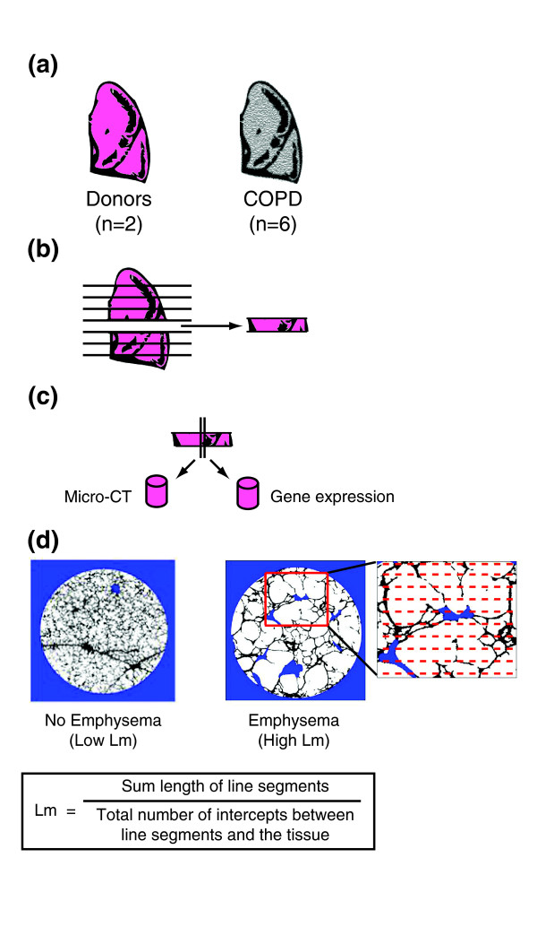
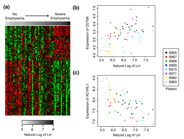
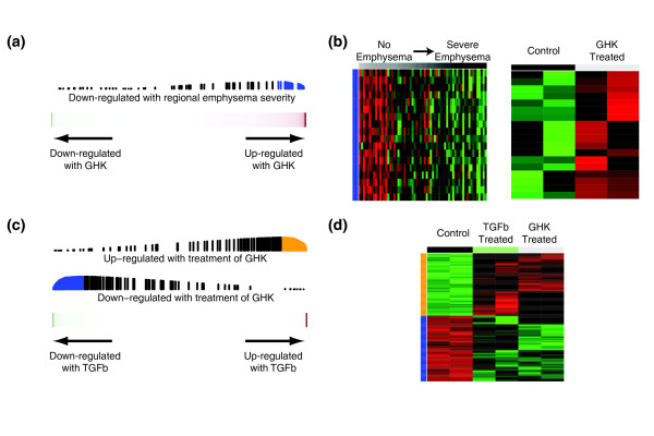
Methods: In order to gain insights into the molecular pathways underlying progression of emphysema and explore computational strategies for identifying COPD therapeutics, we profiled gene expression in lung tissue samples obtained from regions within the same lung with varying amounts of emphysematous destruction from smokers with COPD (8 regions × 8 lungs = 64 samples). Regional emphysema severity was quantified in each tissue sample using the mean linear intercept (Lm) between alveolar walls from micro-CT scans.

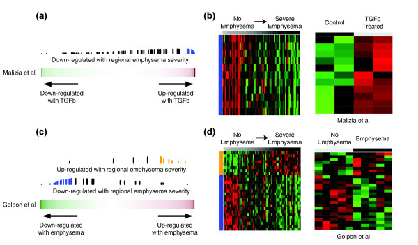
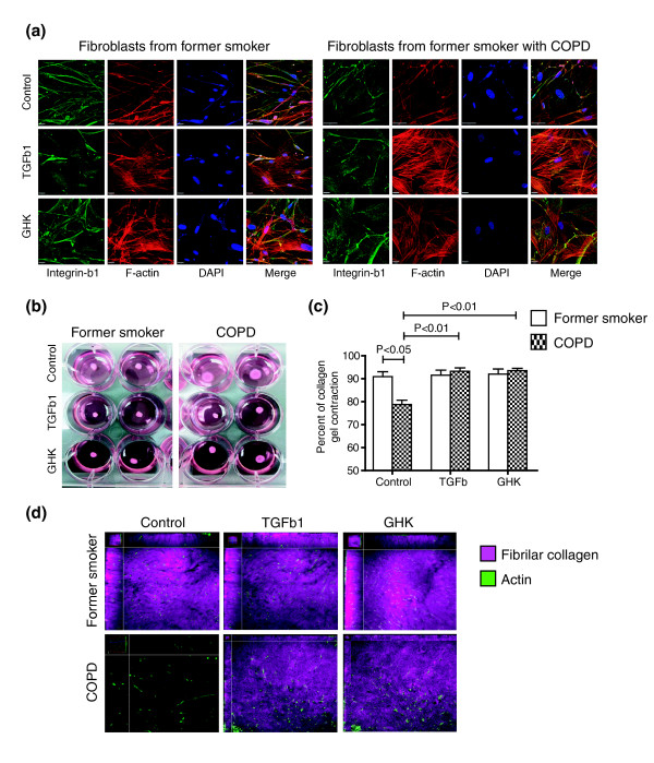
Results: We identified 127 genes whose expression levels were significantly associated with regional emphysema severity while controlling for gene expression differences between individuals. Genes increasing in expression with increasing emphysematous destruction included those involved in inflammation, such as the B-cell receptor signaling pathway, while genes decreasing in expression were enriched in tissue repair processes, including the transforming growth factor beta (TGFβ) pathway, actin organization, and integrin signaling. We found concordant differential expression of these emphysema severity-associated genes in four cross-sectional studies of COPD. Using the Connectivity Map, we identified GHK as a compound that can reverse the gene-expression signature associated with emphysematous destruction and induce expression patterns consistent with TGFβ pathway activation. Treatment of human fibroblasts with GHK recapitulated TGFβ-induced gene-expression patterns, led to the organization of the actin cytoskeleton, and elevated the expression of integrin β1. Furthermore, addition of GHK or TGFβ restored collagen I contraction and remodeling by fibroblasts derived from COPD lungs compared to fibroblasts from former smokers without COPD.

Conclusions: These results demonstrate that gene-expression changes associated with regional emphysema severity within an individual's lung can provide insights into emphysema pathogenesis and identify novel therapeutic opportunities for this deadly disease. They also suggest the need for additional studies to examine the mechanisms by which TGFβ and GHK each reverse the gene-expression signature of emphysematous destruction and the effects of this reversal on disease progression.


Figures

Figure 1
Figure 1
Outline of study design. (a) Whole lungs were removed from patients with severe COPD and from donors, inflated with air, and rapidly frozen in liquid nitrogen vapor. (b) The frozen specimens were cut into 2-cm slices from apex to base of the lung. (c) Adjacent tissue cores were removed from 8 different slices of each lung (8 patients with 8 slices = 64 total regions). (d) Micro-CT was used to measure Lm at 20 evenly spaced intervals throughout one core from each region.
Figure 2
Figure 2
Gene expression signature of regional emphysema severity. (a) Supervised heatmap of genes whose expression is associated with Lm (FDR <0.10). Samples are organized from low to high Lm. Each row corresponds to a gene and each column corresponds to a sample. Green represents lower relative expression and red represents higher relative expression. (b,c) Expression of CD79A (b) and ACVRL1 (c) are plotted against the natural log of Lm with the color of each point indicating the subject from which the sample was derived.
Figure 3
Figure 3
Validation of differential expression for CD79A by immunohistochemistry. Representative images of CD79A-positive cells (arrows) in the alveolar tissue and the small airway walls. Positive staining appears red. Scale = 200 µm; inset = 10 µm.
Figure 4
Figure 4
Relation between gene expression changes associated with regional emphysema severity (Lm) and other gene-expression studies by GSEA. (a) Relation between gene expression changes associated with regional emphysema severity and those induced by TGFβ treatment of A549 cells from Malizia et al.[23]. The color bar represents the fold change between the cell lines treated with and without TGFβ1for 11,910 genes in Malizia et al.[23]. Red indicates a more positive fold change and green indicates a more negative fold change (induced or repressed with TGFβ, respectively). The vertical lines represent the position of genes associated with regional emphysema severity in the ranked gene list. The height of the vertical lines corresponds to the magnitude of the running enrichment score from GSEA. Blue vertical lines indicate that the gene is part of the 'core' enrichment (that is, all the genes from the absolute maximum enrichment score to the end of the ranking). (b) Supervised heatmaps of relative gene expression levels for the core enrichment genes in both the regional emphysema and Malizia et al. datasets (11 genes down-regulated with emphysema severity but up-regulated with TGFβ). Each gene is represented in the same row across heatmaps. (c) Genes changing in expression with increasing regional emphysema severity were enriched in the cross-sectional study of COPD-related gene expression from Golpon et al.[6]. The color bar represents the t-statistic from a t-test between five emphysema patients and five non-smokers for 5,209 genes in Golpon et al.[6]. Blue and orange vertical lines indicate that the gene is part of the core enrichment. (d) Supervised heatmaps of relative gene expression levels for the core enrichment genes in both the regional emphysema and Golpon datasets (8 genes concordantly up-regulated; 19 genes concordantly down-regulated).
Figure 5
Figure 5
Effect of GHK treatment on expression in human lung fibroblasts. (a) Genes decreasing in expression with increasing regional emphysema severity were enriched among genes that are induced by GHK at 10 nM. (b) Supervised heatmaps of relative gene expression levels for the core enrichment genes in both datasets (18 genes down-regulated with Lm but up-regulated with GHK). Each gene is represented in the same row across heatmaps. (c) Genes differentially expressed with treatment of GHK at 0.1 nM were concordantly enriched among genes that change with treatment of TGFβ1. (d) Heatmap of relative gene expression levels for the core enrichment genes (118 genes up-regulated and 124 down-regulated with both GHK and TGFβ1).
Figure 6
Figure 6
Effect of GHK treatment on collagen contraction by fibroblasts from former smokers with COPD. (a) Representative immunofluorescent images of distal lung fibroblasts from former smokers with and without COPD treated with GHK (10 nM), TGFβ1 (10 ng/ml), or media control for 48 h and stained with phalloidin to localize the actin cytoskeleton (red), integrin-β1antibody (green) and DAPI to localize nuclei (blue). (b) Representative images of collagen I gel bioassays at 24 h after being seeded with distal lung fibroblasts from former smokers with and without COPD previously treated with GHK, TGFβ1, or media control for 48 h. (c) The percentage of collagen I contraction was significantly decreased in fibroblasts derived from former smokers with COPD compared to former smokers without COPD (P < 0.05) but was significantly increased with addition of TGFβ1 or GHK (P < 0.01). (d) Representative enface Z-stack slices of three-dimensional reconstructed collagen I gel bioassays demonstrating actin in fibroblasts (green, phalloidin) and second harmonic signal originating from collagen fibrils (purple, 414 nM). Fibroblasts from former smokers with COPD were unable to efficiently remodel collagen into fibrils. However, this intrinsic defect was restored with treatment of TGFβ1 or GHK.

Similar articles

Cited by

References

    1. Miniño AM, Xu J, Kochanek KD, Statistics V. National Vital Statistics Reports Volume 59. NVSS; 2010. Deaths: Preliminary Data for 2008.http://www.cdc.gov/nchs/data/nvsr/nvsr59/nvsr59_02.pdf - PubMed
    1. Lange P, Scharling H, Fabricius P, Vestbo J. Developing COPD: a 25 year follow up study of the general population. Thorax. 2006;4:935–939. doi: 10.1136/thx.2006.062802. - DOI - PMC - PubMed
    1. Park JW, Ryter SW, Choi AMK. Functional significance of apoptosis in chronic obstructive pulmonary disease. COPD. 2007;4:347–353. doi: 10.1080/15412550701603775. - DOI - PubMed
    1. Postma DS, Timens W. Remodeling in asthma and chronic obstructive pulmonary disease. Proc Am Thorac Soc. 2006;4:434–439. doi: 10.1513/pats.200601-006AW. - DOI - PubMed
    1. Rennard SI, Bailey KL. Chronic obstructive pulmonary disease exacerbations: accurate and easy measurement promises much. Am J Respir Crit Care Med. 2012;4:1139–1141. doi: 10.1164/rccm.201202-0227ED. - DOI - PMC - PubMed