

Vectorial secretion of CTGF as a cell-type specific response to LPA and TGF-β in human tubular epithelial cells
- PMID: 22938209
- PMCID: PMC3503564
- DOI: 10.1186/1478-811X-10-25
Vectorial secretion of CTGF as a cell-type specific response to LPA and TGF-β in human tubular epithelial cells
Abstract
Background: Increased expression of the pro-fibrotic protein connective tissue growth factor (CTGF) has been detected in injured kidneys and elevated urinary levels of CTGF are discussed as prognostic marker of chronic kidney disease. There is evidence that epithelial cells lining the renal tubular system contribute to uptake and secretion of CTGF. However, the role of different types of tubular epithelial cells in these processes so far has not been addressed in primary cultures of human cells.
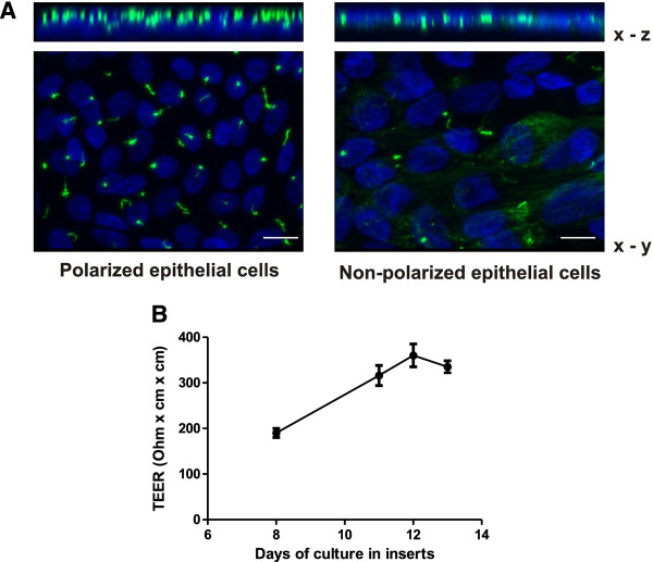
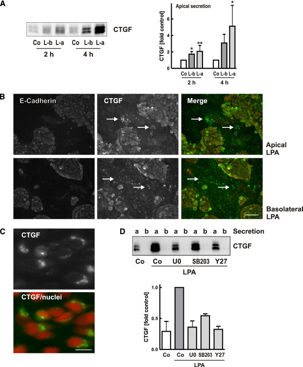
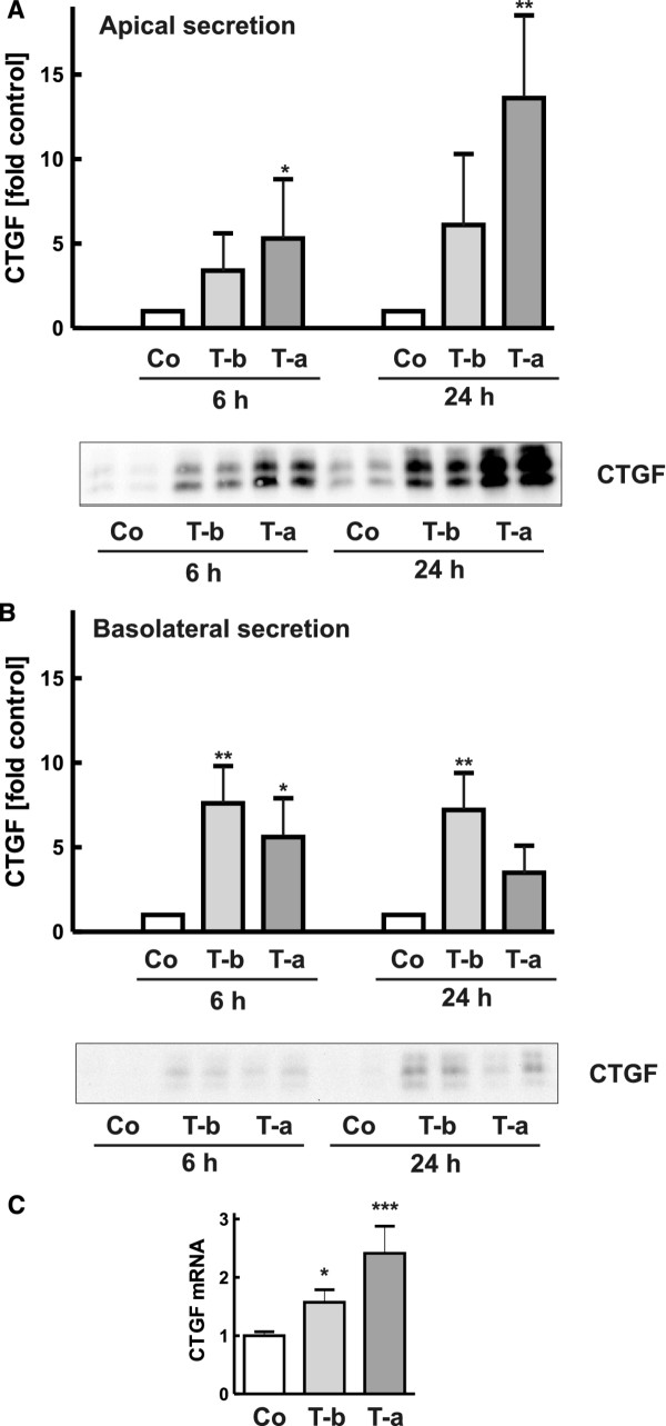
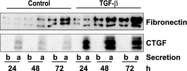
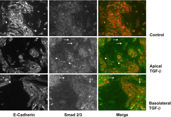
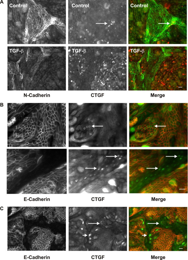
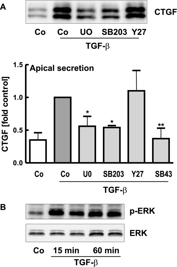
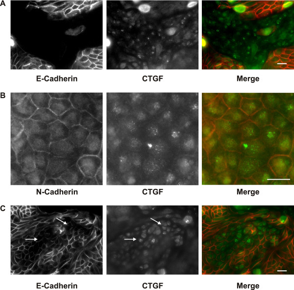
Results: Tubular epithelial cells of proximal and distal origin were isolated from human kidneys and cultured as polarized cells in insert wells. The pro-fibrotic stimuli lysophosphatidic acid (LPA) and transforming growth factor β (TGF-β) were used to induce CTGF secretion.LPA activated CTGF secretion in proximal tubular cells when applied from either the apical or the basolateral side as shown by immunocytochemistry. CTGF was secreted exclusively to the apical side. Signaling pathways activated by LPA included MAP kinase and Rho kinase signaling. TGF-β applied from either side also stimulated CTGF secretion primarily to the apical side with little basolateral release.Interestingly, TGF-β activation induced different signaling pathways depending on the side of TGF-β application. Smad signaling was almost exclusively activated from the basolateral side most prominently in cells of distal origin. Only part of these cells also synthesized CTGF indicating that Smad activation alone was not sufficient for CTGF induction. MAP kinases were involved in apical TGF-β-mediated activation of CTGF synthesis in proximal cells and a subset of epithelial cells of distal origin. This subpopulation of distal tubular cells was also able to internalize recombinant apical CTGF, in addition to proximal cells which were the main cells to take up exogenous CTGF.
Conclusions: Analysis of polarized human primary renal epithelial cells in a transwell system shows that vectorial secretion of the pro-fibrotic protein CTGF depends on the cell type, the stimulus and the signaling pathway activated. In all conditions, CTGF was secreted mainly to the apical side upon TGF-β and LPA treatment and therefore, likely contributes to increased urinary CTGF levels in vivo. Moreover, CTGF secreted basolaterally may be active as paracrine pro-fibrotic mediator.
Figures
Similar articles
-
Connective tissue growth factor induction by lysophosphatidic acid requires transactivation of transforming growth factor type β receptors and the JNK pathway.Cell Signal. 2011 Feb;23(2):449-57. doi: 10.1016/j.cellsig.2010.10.019. Epub 2010 Oct 18. Cell Signal. 2011. PMID: 20965247
-
Exogenous CGRP upregulates profibrogenic growth factors through PKC/JNK signaling pathway in kidney proximal tubular cells.Cell Biol Toxicol. 2018 Aug;34(4):251-262. doi: 10.1007/s10565-017-9399-4. Epub 2017 May 24. Cell Biol Toxicol. 2018. PMID: 28540451
-
Expression of connective tissue growth factor in human renal fibroblasts: regulatory roles of RhoA and cAMP.J Am Soc Nephrol. 2001 Sep;12(9):1853-1861. doi: 10.1681/ASN.V1291853. J Am Soc Nephrol. 2001. PMID: 11518778
-
Lysophosphatidic acid increases proximal tubule cell secretion of profibrotic cytokines PDGF-B and CTGF through LPA2- and Gαq-mediated Rho and αvβ6 integrin-dependent activation of TGF-β.Am J Pathol. 2012 Oct;181(4):1236-49. doi: 10.1016/j.ajpath.2012.06.035. Epub 2012 Aug 10. Am J Pathol. 2012. PMID: 22885106 Free PMC article.
-
[Role of connective tissue growth factor in human renal tubular epithelial cell transdifferentiation in vitro].Zhonghua Yi Xue Za Zhi. 2005 Nov 2;85(41):2920-5. Zhonghua Yi Xue Za Zhi. 2005. PMID: 16324366 Chinese.
Cited by
-
Deficiency of lysophosphatidic acid receptor 3 decreases erythropoietin production in hypoxic mouse kidneys.Lipids Health Dis. 2024 Nov 18;23(1):381. doi: 10.1186/s12944-024-02367-8. Lipids Health Dis. 2024. PMID: 39558335 Free PMC article.
-
Integrated single-nucleus sequencing and spatial architecture analysis identified distinct injured-proximal tubular types in calculi rats.Cell Biosci. 2023 May 19;13(1):92. doi: 10.1186/s13578-023-01041-3. Cell Biosci. 2023. PMID: 37208718 Free PMC article.
-
Proximal tubule LPA1 and LPA2 receptors use divergent signaling pathways to additively increase profibrotic cytokine secretion.Am J Physiol Renal Physiol. 2021 Mar 1;320(3):F359-F374. doi: 10.1152/ajprenal.00494.2020. Epub 2021 Jan 11. Am J Physiol Renal Physiol. 2021. PMID: 33427061 Free PMC article.
-
Metformin and Inhibition of Transforming Growth Factor-Beta Stimulate In Vitro Transport in Primary Renal Tubule Cells.Tissue Eng Part A. 2020 Oct;26(19-20):1091-1098. doi: 10.1089/ten.TEA.2019.0294. Tissue Eng Part A. 2020. PMID: 32312181 Free PMC article.
-
HIF stabilization inhibits renal epithelial cell migration and is associated with cytoskeletal alterations.Sci Rep. 2018 Jun 22;8(1):9497. doi: 10.1038/s41598-018-27918-9. Sci Rep. 2018. PMID: 29934555 Free PMC article.
References
-
- Chen XM, Qi W, Pollock CA. CTGF and chronic kidney fibrosis. Front Biosci (Schol Ed) 2009;1:132–141. - PubMed
-
- Boor P, Floege J. Chronic kidney disease growth factors in renal fibrosis. Clin Exp Pharmacol Physiol. 2011;38:391–400. - PubMed
-
- Tang SC, Leung JC, Lai KN. Diabetic tubulopathy: an emerging entity. Contrib Nephrol. 2011;170:124–134. - PubMed
-
- Wolf G. Renal injury due to renin-angiotensin-aldosterone system activation of the transforming growth factor-beta pathway. Kidney Int. 2006;70:1914–1919. - PubMed
LinkOut - more resources
Full Text Sources
Miscellaneous
