

Prevention and management of limb contractures in neuromuscular diseases
- PMID: 22938881
- PMCID: PMC3482407
- DOI: 10.1016/j.pmr.2012.06.009
Prevention and management of limb contractures in neuromuscular diseases
Abstract
Limb contractures are a common impairment in neuromuscular diseases. They contribute to increased disability from decreased motor performance, mobility limitations, reduced functional range of motion, loss of function for activities of daily living, and increased pain. The pathogenesis of contractures is multifactorial. Myopathic conditions are associated with more severe limb contractures compared with neuropathic disorders. Although the evidence supporting the efficacy of multiple interventions to improve range of motion in neuromuscular diseases in a sustained manner is lacking, there are generally accepted principles with regard to splinting, bracing, stretching, and surgery that help minimize the impact or disability from contractures.
Copyright © 2012 Elsevier Inc. All rights reserved.
Figures
References
-
- Spector SA, Simard CP, Fournier M, Sternlicht E, Edgerton VR. Architectural alterations of rat hind-limb skeletal muscles immobilized at different lengths. Exp Neurol. 1982 Apr;76(1):94–110. - PubMed
-
- Brooke MH, Fenichel GM, Griggs RC, Mendell JR, Moxley R, Florence J, King WM, Pandya S, Robison J, Schierbecker J, et al. Duchenne muscular dystrophy: patterns of clinical progression and effects of supportive therapy. Neurology. 1989 Apr;39(4):475–81. - PubMed
-
- Johnson EW, Zeiter Walter J. Lecture: pathokinesiology of Duchenne muscular dystrophy: implications for management. Arch Phys Med Rehabil. 1977 Jan;58(1):4–7. - PubMed
-
- Sutherland DH, Olshen R, Cooper L, Wyatt M, Leach J, Mubarak S, Schultz P. The pathomechanics of gait in Duchenne muscular dystrophy. Dev Med Child Neurol. 1981 Feb;23(1):3–22. - PubMed
-
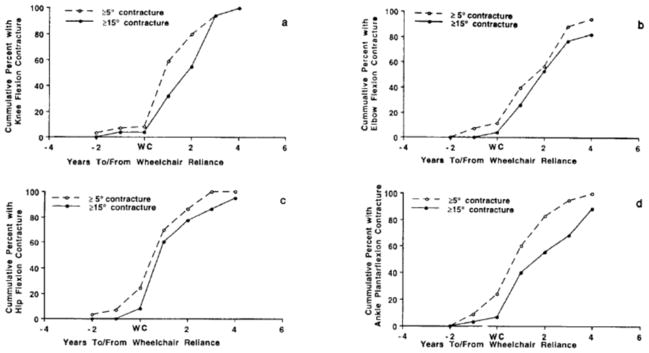
- Archibald KC, Vignos PJ., Jr A study of contractures in muscular dystrophy. Arch Phys Med Rehabil. 1959 Apr;40(4):150–7. - PubMed
Publication types
MeSH terms
Grants and funding
LinkOut - more resources
Full Text Sources
Other Literature Sources
Medical
