. 2012 Sep;18(9):724-33.
doi: 10.1016/j.cardfail.2012.07.005.

Abnormalities of calcium handling proteins in skeletal muscle mirror those of the heart in humans with heart failure: a shared mechanism?

Affiliations

Abnormalities of calcium handling proteins in skeletal muscle mirror those of the heart in humans with heart failure: a shared mechanism?

Holly R Middlekauff et al. J Card Fail. 2012 Sep.

Abstract

Background: In the failing human heart, abnormalities of Ca(2+) cycling have been described, but there is scant knowledge about Ca(2+) handling in the skeletal muscle of humans with heart failure (HF). We tested the hypothesis that in humans with HF, Ca(2+) cycling proteins in skeletal muscle are abnormal.

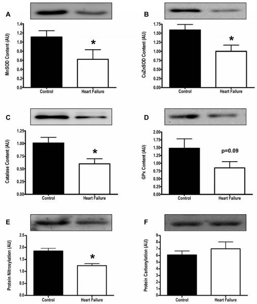
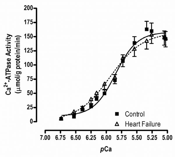
Methods and results: Ten advanced HF patients (50.4 ± 3.7 years), and 9 age-matched controls underwent vastus lateralis biopsy. Western blot analysis showed that sarco(endo)plasmic reticulum Ca(2+)-ATPase (SERCA)2a, which is responsible for Ca(2+) sequestration into the sarcoplasmic reticulum(SR), was lower in HF versus controls (4.8 ± 0.5 vs 7.5 ± 0.8 AU, P = .01). Although phospholamban (PLN), which inhibits SERCA2a, was not different in HF versus controls, phosphorylation (SER16 site) of PLN, which relieves this inhibition, was reduced (0.8 ± 0.1 vs 3.9 ± 0.9 AU, P = .004). Dihydropyridine receptors were reduced in HF, (2.1 ± 0.4 vs 3.6 ± 0.5 AU, P = .04). We tested the hypothesis that these abnormalities of Ca(2+) handling protein content and regulation were due to increased oxidative stress, but oxygen radical scavenger proteins were not elevated in the skeletal muscle of HF patients.

Conclusion: In chronic HF, marked abnormalities of Ca(2+) handling proteins are present in skeletal muscle, which mirror those in failing heart tissue. This suggests a common mechanism, such as chronic augmentation of sympathetic activity and autophosphorylation of Ca(2+)-calmodulin-dependent-protein kinase II.


Figures

Figure 1
Figure 1
Panel A. Individual fiber type percentage for HF patients. The normal range for type 1 fiber type percentage in the vastus lateralis muscle in the UCLA Neuropathy Laboratory is 36-52%, represented on the graph by the hatched area. In 9 of our 10 HF patients, the type 1 fiber percentage was below this normal range, consistent with a fiber shift from type 1 to type 2 fibers. The overall mean type 1 fiber percentage was 30.6±2.8%. HF=heart failure. Panel B. Individual fiber type 1 and 2 diameters, according to sex. The normal range for fiber type 1 and 2 diameter according to sex is represented on the graph by the hatched area. Although mean fiber type 1 and 2 diameters largely fell within the normal range, 30% of HF patients had smaller type1 fiber diameters, and 50% had small type 2 diameters, compared to the normal cut-off values, consistent with muscle atrophy.
Figure 1
Figure 1
Panel A. Individual fiber type percentage for HF patients. The normal range for type 1 fiber type percentage in the vastus lateralis muscle in the UCLA Neuropathy Laboratory is 36-52%, represented on the graph by the hatched area. In 9 of our 10 HF patients, the type 1 fiber percentage was below this normal range, consistent with a fiber shift from type 1 to type 2 fibers. The overall mean type 1 fiber percentage was 30.6±2.8%. HF=heart failure. Panel B. Individual fiber type 1 and 2 diameters, according to sex. The normal range for fiber type 1 and 2 diameter according to sex is represented on the graph by the hatched area. Although mean fiber type 1 and 2 diameters largely fell within the normal range, 30% of HF patients had smaller type1 fiber diameters, and 50% had small type 2 diameters, compared to the normal cut-off values, consistent with muscle atrophy.
Figure 2
Figure 2
Sarcoplasmic reticulum Ca2+ handling protein content. Panels A-C. SERCA1a (fast twitch isoform), CSQ, and RyR content were not different in human vastus lateralis muscle from healthy controls (black bars) and patients with heart failure (white bars). Panels D-F. SERCA2a (slow twitch isoform), DHPR, and the ratio of p-PLN to PLN were all significantly lower in HF patients compared to healthy controls. * p<0.05, SERCA= sarco(endo)plasmic reticulum Ca2+ ATPase , CSQ= calsequestrin , DHPR= dihydropyridine receptor, RyR=ryanodine receptor.
Figure 3
Figure 3
Anti-oxidant protein content. Panels A-D. Anti-oxidant scavengers were not increased, and in fact MnSOD, CuZnSOD and CAT were significantly decreased in HF patients (white bars) compared to healthy controls (black bars). Panel E,F. Prominent bands for carbonylation data revealed no differences in carbonyl groups between controls (black bars) and HF patients (white bars), and protein nitrosylation (prominent bands) was actually lower in HF patients (white bars) versus controls (black bars). *p<0.05, CAT=catalase, GPx=glutathione peroxidase, SOD=superoxide dismutase
Figure 4
Figure 4
Heat shock protein content. Panels A-C. Heat shock proteins (Hsp27, Hsp70, Hsp90) were not increased, and in fact Hsp70 tended to be lower in HF (white bars) vs controls (black bars).
Figure 5
Figure 5
Ca2+-dependent ATPase activity. There was no difference in maximal Ca2+ATPase activity between HF patients and healthy controls (160.1±15.1 vs168.6±14.1 mol/g protein/min, p=0.69). The pCa50, a measure of Ca2+ sensitivity, was not different between HF and controls (5.7±0.08 vs 5.7±0.007nM, p=0.96). However, the Hill coefficient (1.8±0.2 vs 2.4±0.2, p=0.10), an indicator of Ca2+ co-operativity, tended to be lower in HF patients compared to healthy controls.

Similar articles

Cited by

References

    1. Franciosa JA, Park M, Levine TB. Lack of correlation between exercise capacity and indices of resting left ventricular performance in heart failure. Am J Cardiol. 1991;47:33–9. - PubMed
    1. Higginbotham M, Morris KG, Conn EH, Coleman RE, Cobb FR. Determinants of variable exercise performance among patients with severe left ventricular dysfunction. Am J Cardiol. 1983;51:51–2. - PubMed
    1. Maskin CS, Forman R, Sonnenblick EH, Frishman WH, LeJemtel TH. Failure of dobutamine to increase exercise capacity despite hemodynamic improvement in severe chronic heart failure. Am J Cardiol. 1983;51:177–82. - PubMed
    1. Middlekauff HR. Making the case for skeletal myopathy as the major limitation of exercise capacity in heart failure. Circ Heart Fail. 2010;3:537–46. - PMC - PubMed
    1. Harrington D, Anker SD, Chua TP, Webb-Peploe KM, Ponikowski PP, Poole-Wilson PA, et al. Skeletal muscle function and its relation to exercise tolerance in chronic HF. J Am Coll Cardiol. 1997;30:1758–64. - PubMed

Substances