

Optical coherence tomography grading reproducibility during the Comparison of Age-related Macular Degeneration Treatments Trials
- PMID: 22939114
- PMCID: PMC3631582
- DOI: 10.1016/j.ophtha.2012.06.040
Optical coherence tomography grading reproducibility during the Comparison of Age-related Macular Degeneration Treatments Trials
Abstract
Objective: To report reading center reproducibility during grading of Stratus optical coherence tomography (OCT) (Carl Zeiss Meditec, Dublin, CA) images obtained during the Comparison of Age-Related Macular Degeneration Treatments Trials (CATT).
Design: Prospective, clinical trial.
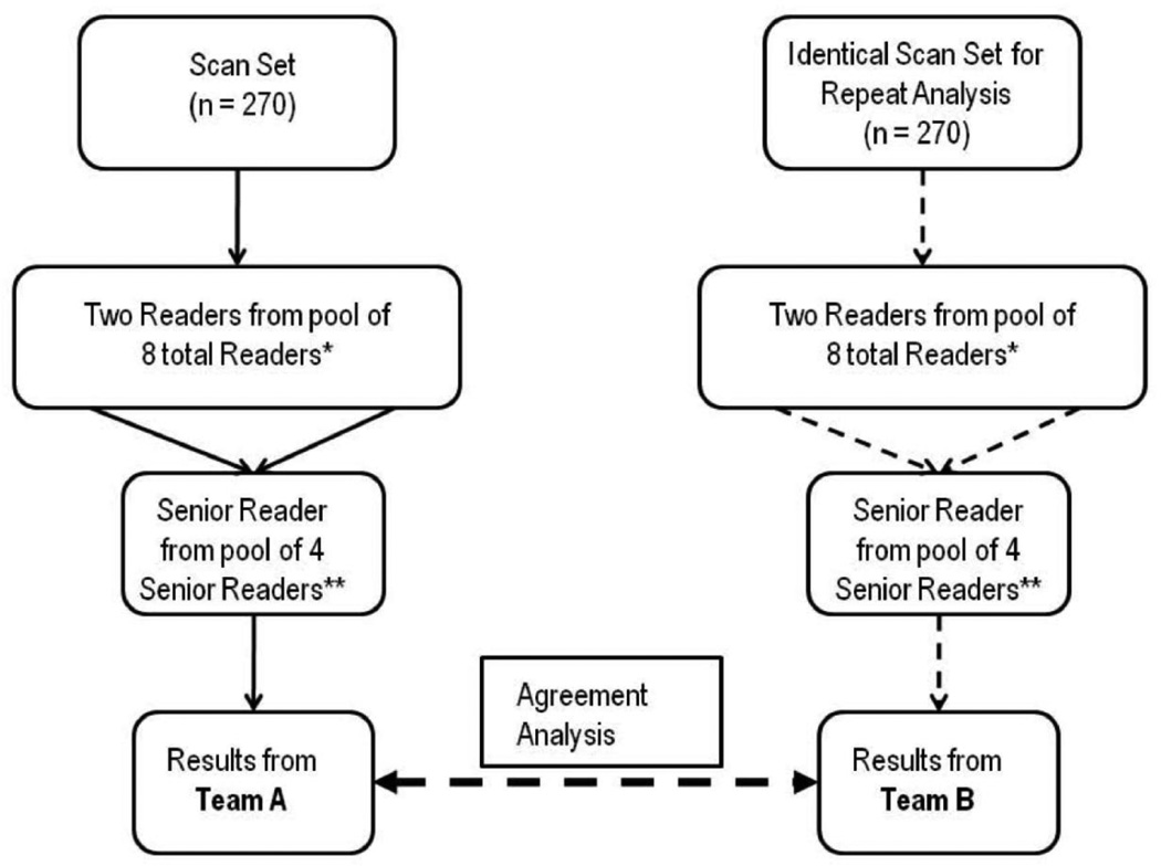
Participants: Independent reading teams reevaluated 270 OCT scans randomly sampled from the first 2 years of CATT enrollment. To assess temporal drift, a cohort of 23 scans submitted during the initial portion of the CATT study was longitudinally followed with serial reproducibility analysis.
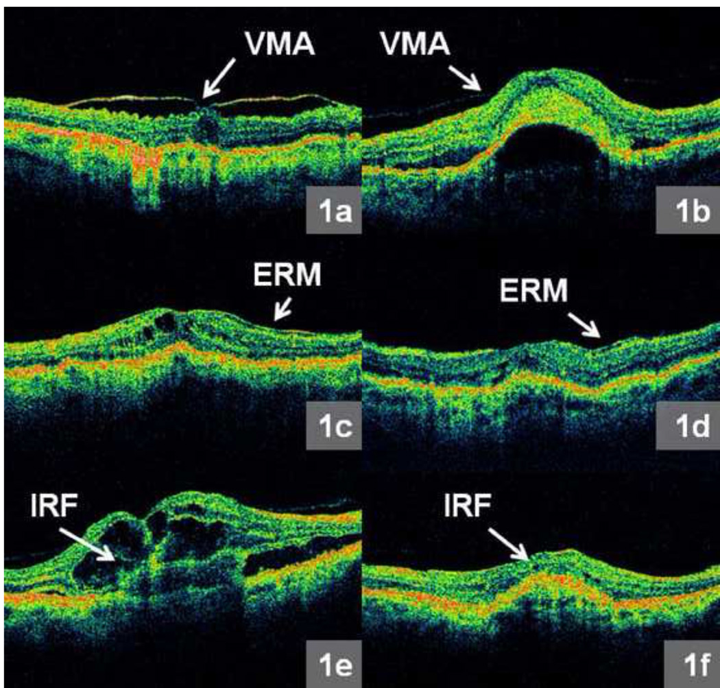
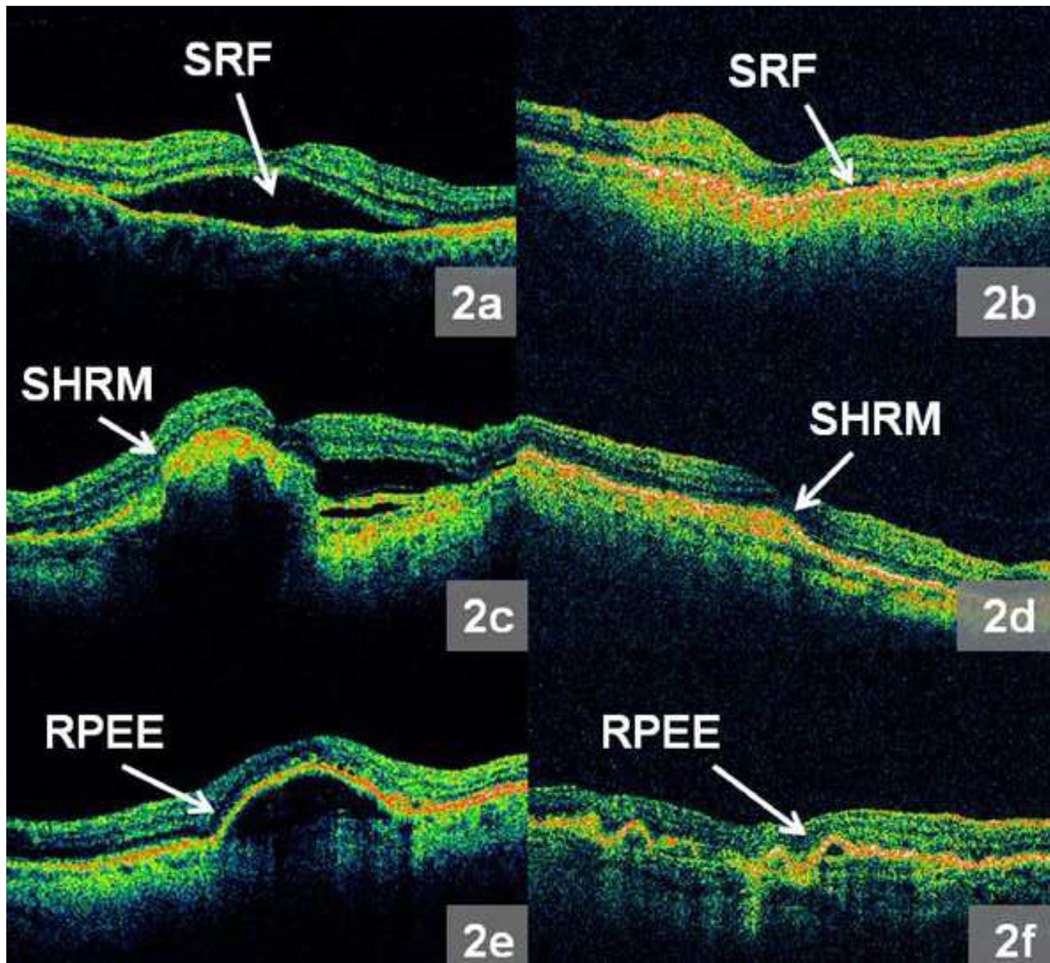
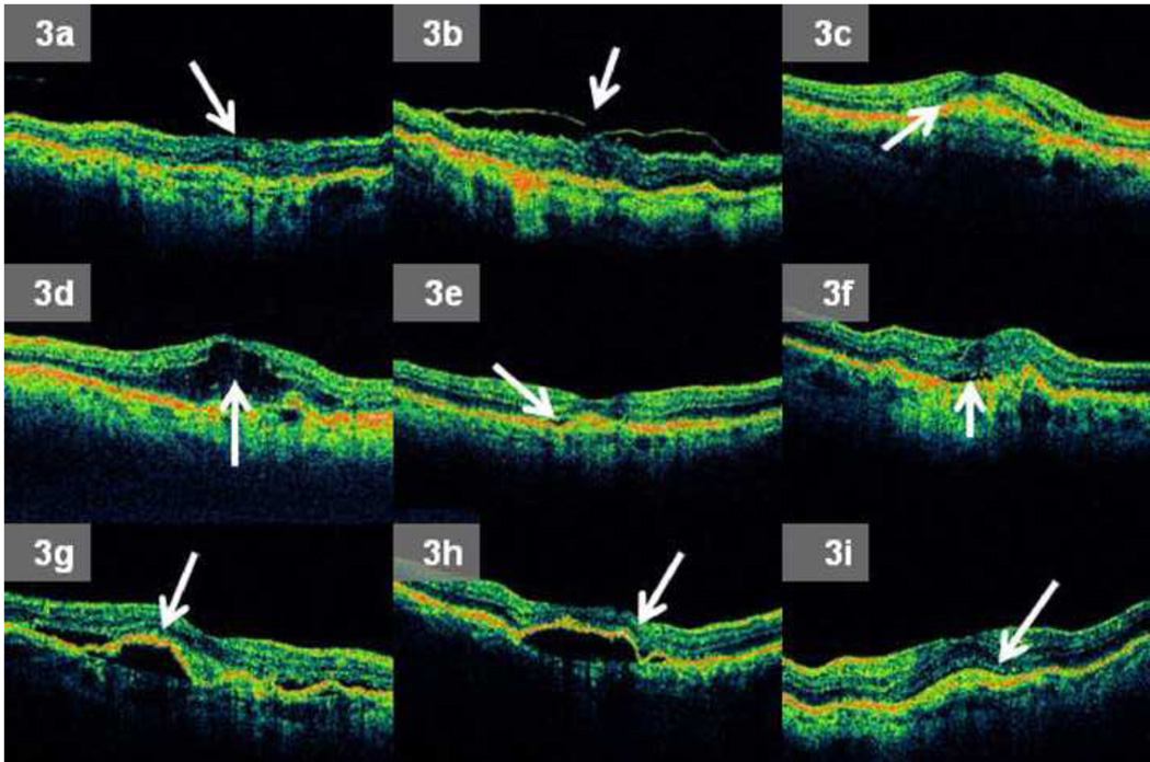
Intervention: The CATT readers performed standardized grading of OCT images. A reader team, composed of 2 independent readers and a senior reader, evaluated each scan. Grading included the CATT OCT end points of total thickness at the foveal center point and intraretinal fluid (IRF), subretinal fluid (SRF), and subretinal pigment epithelium (RPE) fluid. Independent reading teams masked to the results of initial grading reevaluated scans to determine the reproducibility of qualitative grading and measurements.
Main outcome measures: Categorical grading agreement was reported using percent agreement and kappa statistic, and measurement agreement was reported using intraclass correlations and paired differences.
Results: Reading center teams reproducibly graded IRF (percent agreement = 73%, kappa = 0.48; 95% confidence interval [CI], 0.38-0.58), SRF (percent agreement = 90%; kappa = 0.80; 95% CI, 0.73-0.87), and sub-RPE fluid (percent agreement 88%; kappa = 0.75; 95% CI, 0.67-0.83). For independent reading center team measurements of total thickness at the foveal center point, the intraclass correlation was 0.99 (95% CI, 0.99-0.99), and the mean paired difference between reading center teams was 4 μm (95% limits of agreement, -55 to 47 μm). There was no qualitative or quantitative grading drift.
Conclusions: The standardized protocols used to evaluate OCT scans from the CATT study were reproducible. The methods used are suitable to monitor OCT imaging data from a large, neovascular age-related macular degeneration, interventional, multicenter study.
Financial disclosure(s): The author(s) have no proprietary or commercial interest in any materials discussed in this article.
Copyright © 2012 American Academy of Ophthalmology. Published by Elsevier Inc. All rights reserved.
Conflict of interest statement
Conflict of Interest: No conflicting relationship exists for any author.
Figures

References
-
- Hee MR, Baumal CR, Puliafito CA, et al. Optical coherence tomography of age-related macular degeneration and choroidal neovascularization. Ophthalmology. 1996;103:1260–1270. - PubMed
-
- Jaffe GJ, Caprioli J. Optical coherence tomography to detect and manage retinal disease and glaucoma. Am J Ophthalmol. 2004;137:156–169. - PubMed
-
- Ting TD, Oh M, Cox TA, et al. Decreased visual acuity associated with cystoid macular edema in neovascular age-related macular degeneration. Arch Ophthalmol. 2002;120:731–737. - PubMed
-
- Hee MR, Puliafito CA, Wong C, et al. Quantitative assessment of macular edema with optical coherence tomography. Arch Ophthalmol. 1995;113:1019–1029. - PubMed
Publication types
MeSH terms
Substances
Grants and funding
LinkOut - more resources
Full Text Sources
Medical
Miscellaneous
