Clinical neuropathology practice guide 5-2012: updated guideline for the diagnosis of antineuronal antibodies
- PMID: 22939174
- PMCID: PMC3663458
- DOI: 10.5414/np300545
Clinical neuropathology practice guide 5-2012: updated guideline for the diagnosis of antineuronal antibodies
Abstract
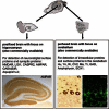
In recent years there is an increasing description of novel anti-neuronal antibodies that are associated with paraneoplastic and non-paraneoplastic neurological syndromes. These antibodies are useful in clinical practice to confirm the immunmediated origin of the neurological disorder and are helpful in tumor search. Currently, antineuronal antibodies can be classified according to the location of the recognized antigen into two groups, 1.) intraneuronal antigens and 2.) antigens located in the cell membrane. Different techniques are established for detecting these antibodies: tissue-based assay (TBA), cell-based assay (CBA), immunoblot, immunoprecipitation assay (IP), and ELISA. TBA detect most of the antibodies, however, different pretreatment methods of rat brain are necessary to visualize either Group 1 or 2 antibodies. Higher specificity is provided by immunoblots, applicable for Group 1 antibodies, and CBA, suitable for Group 2 antibodies. IP and ELISA may be useful for the detection of specific antibodies or to solve particular issues such as antibody titers. Diagnosis of paraneoplastic and non-paraneoplastic neurological syndromes has important implications on treatment and follow-up of patients. Selection and proper combination of test systems and appropriate knowledge of the clinical information will provide a maximum of sensitivity and specificity in identifying the associated antibody.
Figures
References
-
- Graus F Saiz A Dalmau J Antibodies and neuronal autoimmune disorders of the CNS. J Neurol. 2010; 257: 509–517 doi:10.1007/s00415-009-5431-9 - PubMed
-
- de Graaff E Maat P Hulsenboom E van den Berg R van den Bent M Demmers J Lugtenburg PJ Hoogenraad CC Sillevis Smitt P Identification of delta/notch-like epidermal growth factor-related receptor as the Tr antigen in paraneoplastic cerebellar degeneration. Ann Neurol. 2012; 71: 815–824 doi:10.1002/ana.23550 - PubMed
-
- Johannis W Renno JH Wielckens K Voltz R Ma2 antibodies: an evaluation of commercially available detection methods. Clin Lab. 2011; 57: 321–326 - PubMed
Publication types
MeSH terms
Substances
Grants and funding
LinkOut - more resources
Full Text Sources
Other Literature Sources
Medical


