. 2012 Dec;16(8):1550-64.
doi: 10.1016/j.media.2012.07.004. Epub 2012 Aug 9.

Reconstruction of fetal brain MRI with intensity matching and complete outlier removal

Affiliations

Reconstruction of fetal brain MRI with intensity matching and complete outlier removal

Maria Kuklisova-Murgasova et al. Med Image Anal. 2012 Dec.

Abstract

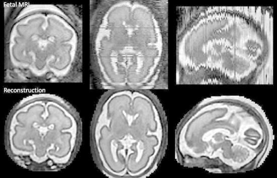
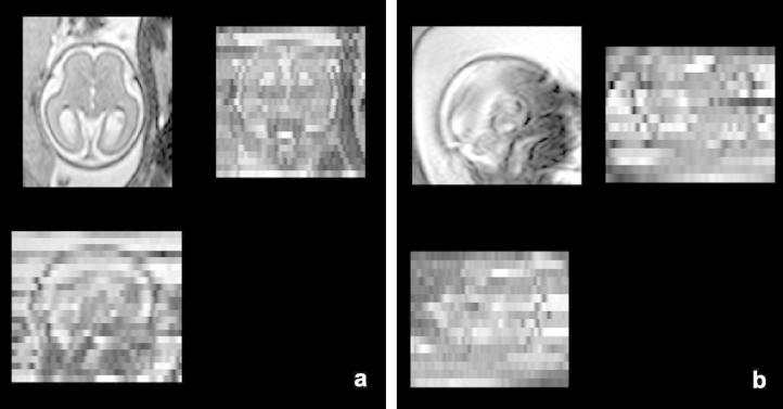
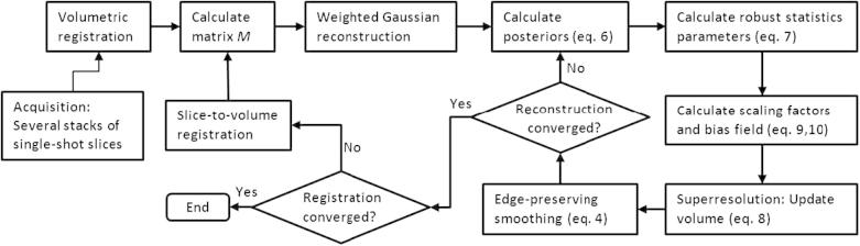
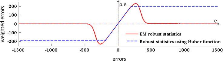
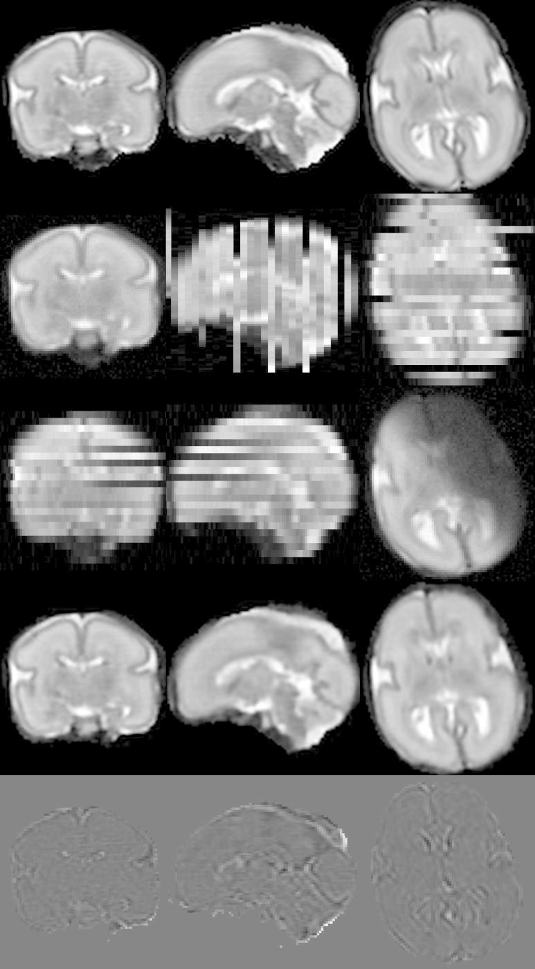
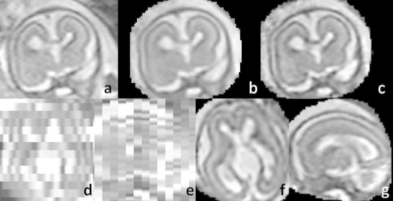
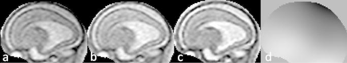
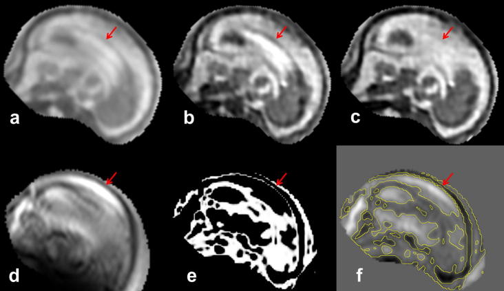
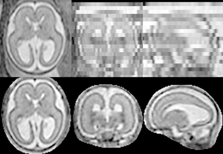
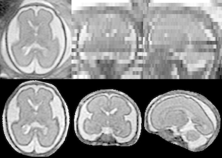
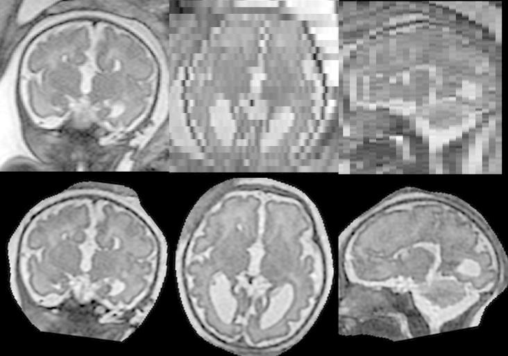
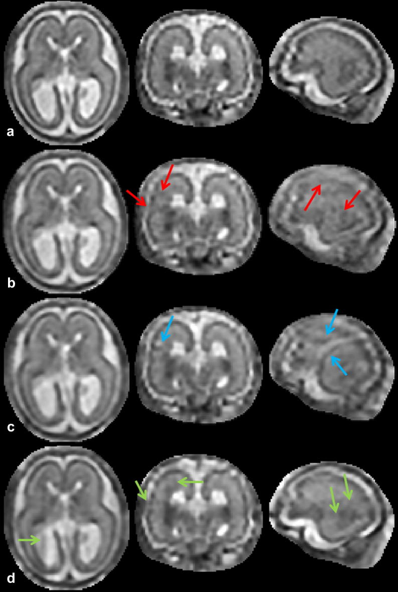
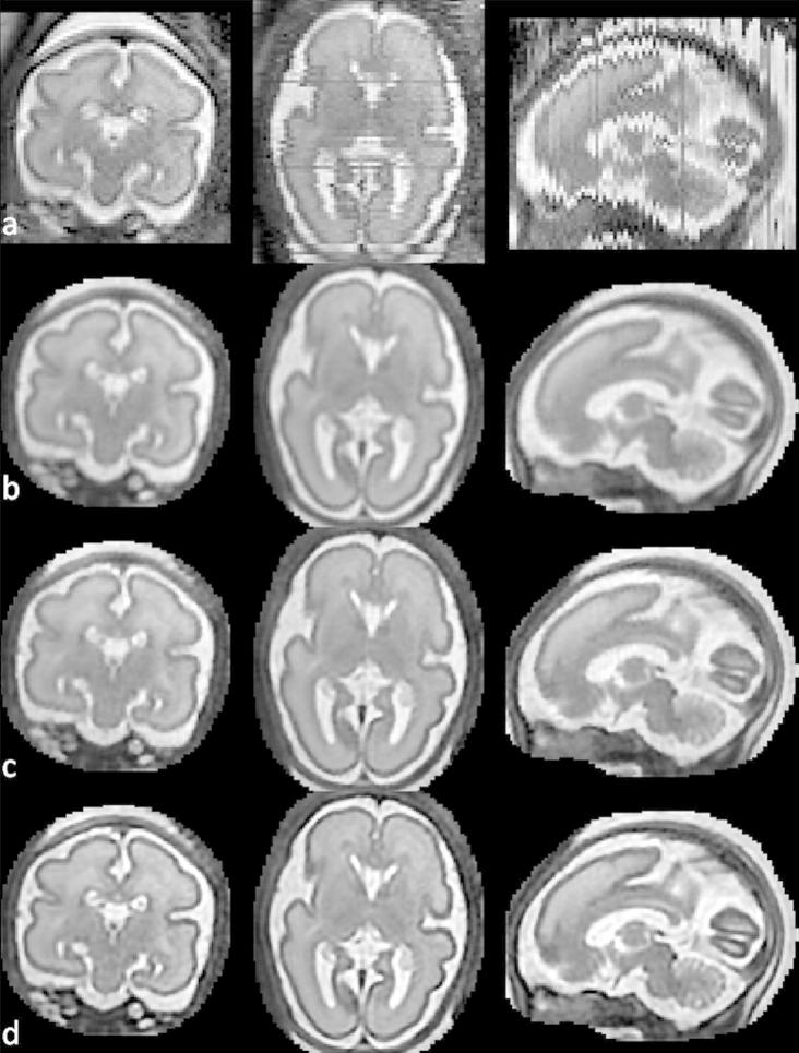
We propose a method for the reconstruction of volumetric fetal MRI from 2D slices, comprising super-resolution reconstruction of the volume interleaved with slice-to-volume registration to correct for the motion. The method incorporates novel intensity matching of acquired 2D slices and robust statistics which completely excludes identified misregistered or corrupted voxels and slices. The reconstruction method is applied to motion-corrupted data simulated from MRI of a preterm neonate, as well as 10 clinically acquired thick-slice fetal MRI scans and three scan-sequence optimized thin-slice fetal datasets. The proposed method produced high quality reconstruction results from all the datasets to which it was applied. Quantitative analysis performed on simulated and clinical data shows that both intensity matching and robust statistics result in statistically significant improvement of super-resolution reconstruction. The proposed novel EM-based robust statistics also improves the reconstruction when compared to previously proposed Huber robust statistics. The best results are obtained when thin-slice data and the correct approximation of the point spread function is used. This paper addresses the need for a comprehensive reconstruction algorithm of 3D fetal MRI, so far lacking in the scientific literature.


Figures

None
Graphical abstract
Fig. 1
Fig. 1
Motion artifacts in fetal MRI shown in three orthogonal plane views. (a) A stack acquired in axial direction exhibiting a smaller amount of motion. The individual images are of high quality but do not form a consistent representation. Structures can still be recognized in the through-plane direction. (b) A stack acquired in sagittal direction with heavy motion. Some slices are corrupted by motion artifacts. Structures cannot be recognized in the through-plane direction.
Fig. 2
Fig. 2
Overview of the proposed methodology.
Fig. 3
Fig. 3
Influence functions of EM and Huber robust statistics. During an iteration of the super-resolution algorithm, errors ejk are redistributed to update the reconstructed volume (Eq. (4)). If robust statistics are used, values pjkejk are redistributed instead (Eq. (8)). Robust EM statistics transform large errors to values close to zero (red line) while the Huber function only thresholds the error values at a certain value (dashed blue line). The x-axis represents error values ejk, and the y-axis represents error values after applying robust statistics. (For interpretation of the references to color in this figure legend, the reader is referred to the web version of this article.)
Fig. 4
Fig. 4
Simulation of fetal brain MRI from a neonatal subject with GA 27 weeks. First row: Neonatal volume. Second row: Simulated coronal stack with 6 slices with large displacement to simulate misregistered outliers. Third row: Simulated transversal stack with three corrupted slices. Fourth row: Reconstruction using six stacks, which include stacks shown in the second and third row, demonstrates the good performance of the method when compared to neonatal volume int the first row. Fifth row: The difference between original and reconstructed image.
Fig. 5
Fig. 5
Comparison of slice weights using (a) EM and (b) Huber robust statistics, plotted against TRE calculated for each slice. Corrupted slices are shown as red circles and the slices which have been deliberately assigned large displacements during simulation are denoted by green crosses. Black asterisks denote slices with small region of interest with little information to guide registration towards correct alignment. The corrupted and misplaced slices are completely removed using EM robust statistics (zero weights), while their weight is only reduced when Huber robust statistics are used. (For interpretation of the references to color in this figure legend, the reader is referred to the web version of this article.)
Fig. 6
Fig. 6
Brain MRI of a 20 week old fetus reconstructed from only 64 slices of 4 mm thickness using the proposed method. First row: (a) An acquired slice. (b) The same slice simulated from the reconstructed volume. (c) Corresponding plane from the reconstructed volume. Second row: Axial and transversal planes orthogonal to the acquired slice shown in (a): original stacks (d and e) and the reconstructed volume (f and g).
Fig. 7
Fig. 7
An example of the bias field estimated during reconstruction of the 23 week old fetus: (a) An acquired slice. (b) The scaled and bias-corrected slice. (c) The corresponding plane of the reconstructed volume. (d) The estimated bias field.
Fig. 8
Fig. 8
Artifacts in the reconstructed volume of the 23 weeks old fetus caused by a corrupted slice. (a) Initialization using Gaussian weighted reconstruction (Rousseau et al., 2006). (b) Reconstruction without robust statistics. (c) Reconstruction with EM robust statistics. (d) The corrupted slice. (e) The posteriors pjk. (h) The error between the corrupted slice and the corresponding simulated slice with an overlaid 0.5 isoline of the posteriors. The red arrow points to the skull in the misaligned corrupted slice, which appears as an artifact in the reconstructions if no robust statistics are used. (For interpretation of the references to color in this figure legend, the reader is referred to the web version of this article.)
Fig. 9
Fig. 9
Reconstructed fetal brain MRI of a subject with GA 23 weeks using only five stacks of slices. First row: One of the acquired stacks. Second row: Reconstructed volume.
Fig. 10
Fig. 10
Reconstructed fetal brain MRI of a subject with GA 26 weeks using only five stacks of slices. First row: One of the acquired stacks. Second row: Reconstructed volume.
Fig. 11
Fig. 11
Reconstructed fetal brain MRI of a subject with GA 34 weeks using only five stacks of slices. First row: One of the acquired stacks. Second row: Reconstructed volume.
Fig. 12
Fig. 12
Reconstructions of the 23 week old fetus from four stacks using four methods evaluated in Table 6: (a) Reconstruction with EM robust statistics and intensity matching (Full). (b) Reconstruction with Huber robust statistics and intensity matching (Huber). (c) Reconstruction with no robust statistics and intensity matching (No robust). (d) Reconstruction with EM robust statistics and no intensity matching (No matching). Red arrows in column (b) show artifacts caused by incomplete removal of misregistered or corrupted slices when Huber robust statistics are used. Blue arrows in column (c) point to artifacts caused by misregistered or corrupted slices when no robust statistics is used. Green arrows show artifacts of intensity inconsistencies in column (d). Compare to Fig. 9 where all five stacks of the same dataset were used for reconstruction using our method. (For interpretation of the references to color in this figure legend, the reader is referred to the web version of this article.)
Fig. 13
Fig. 13
Reconstructed fetal brain MRI of a subject with GA 28 weeks using eight stacks of data acquired using optimized sequences. (a) One of the acquired stacks. (b) Reconstruction using multilevel B-splines. (c) Super-resolution reconstruction with PSF derived from the voxel spacing (FWHM 1.176 mm in-plane and 1.25 mm through-plane). (d) Super-resolution reconstruction with PSF matched to the data (FWHM 1.4 mm in-plane and 2.5 mm through-plane). The same motion correction parameters, determined by application of the full algorithm, were employed in all cases, with only the final reconstruction being performed using the three different reconstruction approaches. Intensity matching and robust statistics were used during all three reconstructions.

Similar articles

Cited by

References

    1. Ashburner J., Friston K.J. Unified segmentation. NeuroImage. 2005;26:839–851. - PubMed
    1. Bertelsen, A., Aljabar, P., Xue, H., Srinivasan, L., Hayat, T., Allsop, J., Rueckert, D., Rutherford, M.R., Hajnal, J.V., 2009. Improved slice to volume reconstruction of the fetal brain for automated cortex segmentation. In: Proceedings of the International Society for, Magnetic Resonance in Medicine, p. 3437.
    1. Charbonnier P., Blanc-Feraud L., Aubert G., Barlaud M. Deterministic edge-preserving regularization in computed imaging. IEEE Transactions on Image Processing. 1997;6:298–311. - PubMed
    1. Clouchoux C., Kudelski D., Gholipour A., Warfield S., Viseur S., Bouyssi-Kobar M., Mari J.L., Evans A., du Plessis A., Limperopoulos C. Quantitative in vivo MRI measurement of cortical development in the fetus. Brain Structure and Function. 2012;217:127–139. - PubMed
    1. Damodaram M., Story L., Allsop J., McGuinness A., Patel A., Kumar S., Rutherford M. 3-dimensional MR reconstruction and brain volumetry in IUGR fetuses. International Journal of Gynecology and Obstetrics. 2009;107:S463–S464. (Abstracts of XIX FIGO World Congress of Gynecology and Obstetrics)