Review
. 2012 Jan;22(1):1-6.
doi: 10.1016/j.tcm.2012.05.017.

Translational findings from cardiovascular stem cell research

Affiliations
Review

Translational findings from cardiovascular stem cell research

Ramesh Mazhari et al. Trends Cardiovasc Med. 2012 Jan.

Abstract

The possibility of using stem cells to regenerate damaged myocardium has been actively investigated since the late 1990s. Consistent with the traditional view that the heart is a "postmitotic" organ that possesses minimal capacity for self-repair, much of the preclinical and clinical work has focused exclusively on introducing stem cells into the heart, with the hope of differentiation of these cells into functioning cardiomyocytes. This approach is ongoing and retains promise but to date has yielded inconsistent successes. More recently, it has become widely appreciated that the heart possesses endogenous repair mechanisms that, if adequately stimulated, might regenerate damaged cardiac tissue from in situ cardiac stem cells. Accordingly, much recent work has focused on engaging and enhancing endogenous cardiac repair mechanisms. This article reviews the literature on stem cell-based myocardial regeneration, placing emphasis on the mutually enriching interaction between basic and clinical research.


Figures

Figure 1
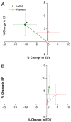
Figure 1. Impact of human MSC treatment on prevention of LV remodeling following myocardial infarction, with permission from Hare et al., JACC 2009; 54(24): 2277-2286
Changes in left ventricular (LV) ejection fraction (EF) are plotted against the changes in LV end-systolic volume (ESV) (A) and end-diastolic volume (EDV) (B) during follow-up. Human mesenchymal stem cell (hMSC) patients (n = 21 at 3 months, n = 18 at 6 and 12 months) exhibit evidence of attenuated remodeling with no increase in LV EDV and a decline in LV ESV, whereas placebo patients (n = 13 at 3 months, n = 11 at 6 and 12 months) demonstrate evidence of LV chamber enlargement consistent with progressive ventricular remodeling. *p = 0.005 versus baseline.
Figure 2
Figure 2. Echocardiographic analysis of CSC-treated patients and controls in SCIPIO trial, with permission from Bolli et al., Lancet 2011; 378(9806): 1847-1857
(A) Left ventricular ejection fraction (measured by use of three-dimensional echocardiography) at 4 months after baseline in control and CSC-treated patients. (B) Ejection fraction at 4 months and 12 months after baseline in the CSC-treated patients who had 1 year of follow-up. (C) Change in ejection fraction from baseline at 4 months and 12 months in CSC-treated patients. (D) Wall motion score index at 4 months after baseline in control and CSC-treated patients. (E) Wall motion score index at 4 months and 12 months after baseline in the CSC-treated patients who had 1 year of follow-up. Boxes represent the mean values and (error bars represent SE). P values are reported for difference between baseline and 4 months and between baseline and 12 months. CSC=cardiac stem cell. As shown, CSC-treated patients had improvement in the regional wall motion score index, and overall ejection fraction at 4 and 12 months following treatment.
Figure 3
Figure 3. MSCs stimulate endogenous CSCs, with permission from Hatzistergos et al. Circulation Research 2010;107:913-922
(A) Graph depicting the contribution of cardiomyocyte precursors following exogenous administration of MSCs (green line) and endogenous CSCs (orange line) during cardiac repair after MI. At 2 weeks, MSCs activate endogenous expansion of c-kit+ CSCs (orange line) (B) Two weeks following TEI, the number of C-kit+ cells co-expressing GATA-4 is greater in MSCs versus non-MSCs treated hearts. (C and D) The 2-week-old chimeric myocardium contains mature cardiomyocytes (open arrow), immature MSCs (inset), and cardiac precursors of MSCs origin (arrow), coupled to host myocardium by connexin-43 gap junctions. Interestingly, endogenous c-kit+ CSCs are found in close proximity to MSCs (D). (E) Cluster of c-kit+ CSCs in an MSC-treated heart; numerous CSCs are committed to cardiac lineage documented by GATA-4 and MDR-1 co-expression (arrows). (F) Few isolated c-kit+ cells were found in non-MSC-treated animals.

References

    1. Abdel-Latif A, Bolli R, Tleyjeh et al. Adult bone marrow-derived cells for cardiac repair: a systematic review and meta-analysis. Archives of internal medicine. 2007;167(10):989–997. doi: 10.1001/archinte.167.10.989. Research Support, N.I.H., ExtramuralReview. - DOI - PubMed
    1. Amado LC, Saliaris AP, Schuleri KH, et al. Cardiac repair with intramyocardial injection of allogeneic mesenchymal stem cells after myocardial infarction. Proceedings of the National Academy of Sciences of the United States of America. 2005;102(32):11474–11479. doi: 10.1073/pnas.0504388102. Comparative StudyResearch Support, N.I.H., ExtramuralResearch Support, Non-U.S. Gov’t. - DOI - PMC - PubMed
    1. Anversa P, Nadal-Ginard B. Myocyte renewal and ventricular remodelling. Nature. 2002;415(6868):240–243. doi: 10.1038/415240a. Research Support, U.S. Gov’t, P.H.S.Review. - DOI - PubMed
    1. Assmus B, Rolf A, Erbs S, et al. Clinical outcome 2 years after intracoronary administration of bone marrow-derived progenitor cells in acute myocardial infarction. Circulation Heart failure. 2010a;3(1):89–96. doi: 10.1161/CIRCHEARTFAILURE.108.843243. Multicenter StudyRandomized Controlled Trial Research Support, Non-U.S. Gov’t. - DOI - PubMed
    1. Assmus B, Rolf A, Erbs S, et al. Clinical outcome 2 years after intracoronary administration of bone marrow-derived progenitor cells in acute myocardial infarction. Circ Heart Fail. 2010b;3(1):89–96. doi: 10.1161/CIRCHEARTFAILURE.108.843243.CIRCHEARTFAILURE.108.843243 - DOI - PubMed

Publication types