. 2012 Sep 21;37(3):549-62.
doi: 10.1016/j.immuni.2012.05.029. Epub 2012 Aug 30.

Inflammatory monocytes activate memory CD8(+) T and innate NK lymphocytes independent of cognate antigen during microbial pathogen invasion

Affiliations

Inflammatory monocytes activate memory CD8(+) T and innate NK lymphocytes independent of cognate antigen during microbial pathogen invasion

Saïdi M'Homa Soudja et al. Immunity. .

Abstract

Memory CD8(+) T cells induced upon immunization exhibit improved functional features that contribute to protection of immunized hosts. Although both cognate antigen recognition and inflammation are important for memory CD8(+) T cell reactivation, the relative contribution of these factors and the cell types providing these signals in vivo are poorly defined. Here, we show that Ly6C(+)CCR2(+) inflammatory monocytes, a subset of monocytes, largely orchestrate memory CD8(+) T and NK lymphocytes activation by differentiating into interleukin-18 (IL-18)- and IL-15-producing cells in an inflammasome and type I interferon-IRF3-dependent manner. Memory CD8(+) T cells became potent effector cells by sensing inflammation from monocytes independently of their cognate antigen. Like NK cells, they underwent rapid mobilization, upregulated intense and sustained effector functions during bacterial, viral, and parasitic infections, and contributed to innate responses and protection in vivo. Thus, inflammatory monocyte-derived IL-18 and IL-15 are critical to initiate memory CD8(+) T and NK lymphocytes differentiation into antimicrobial effector cells.


Conflict of interest statement

Author Information

The authors declare that no competing interests exist.

Figures

Figure 1
Figure 1. Early activation of memory CD8+ T cells is independent of cognate antigen recognition
To generate memory CD8+ T cells, wild-type (WT) C57BL/6 (B6) mice adoptively transferred with 500 OT-I GFP+ cells (A–C) or BALB/c (D) mice were then immunized with WT Lm-Ova or WT Lm (BALB/c) and challenged 6 weeks (wks) later with WT Lm-Ova (blue), Lm (black), Lm-LLOser92 (D, dark blue) or left unchallenged (grey); labeled as “memory”. For “naïve” CD8+ T cells, 3×10⁶ naïve OT-I GFP+ cells were transferred into WT B6 mice. At indicated times (A) or 8 hrs later (B–D), spleens cell suspensions incubated with Golgi Plug were stained for cell surface or functional intracellular markers as specified on the graphs. In (D), endogenous memory CD8+ T cells were tracked using LLO91–99-H-2Kd tetramers (tet). Data show expression of these markers after gating on memory (OT-I, endogenous) CD8+ T cells, with the relative proportion (%) and expression levels (Mean Fluorescence intensity, MFI) of memory cells. Representative flow cytometry histograms are shown. Data pool 3–4 independent replicate experiments (n=8–10 mice) with p-values.
Figure 2
Figure 2. Memory CD8+ T cells contribute to innate immune responses and protection
WT B6 mice transferred with OT-I GFP+ cells were immunized with WT Lm-Ova (A–F) or left unimmunized (D, E). Animals were challenged 6 wks later with WT Lm (A, F), MCMV (B, D) or Streptococcus pneumoniae (C, E). In (G, H) mice were immunized with the lymphochorionmeningitis virus (LCMV). Sixty days later, animals were challenged with WT Lm (G) or LCMV-specific H2-Db/GP33–41 and H2-Db-NP396–404 memory CD8+ T cells were purified and transferred into naïve B6 mice subsequently challenged with WT Lm (H). Spleens (A, B, D, G) and lungs (C, E) were harvested at indicated times, incubated with Golgi Plug and stained for CD8, CD3, LCMV tetramers and NKp46 (NK cell) cell surface markers and intracellular IFN-γ and GrB. In (A–C, G), data show the relative proportion (%) and fold increase in MFI (activated to resting cells) of IFN-γ+ or GrB+ cells amongst OT-I (black), NK (red) cells, and LCMV tet+ cells after gating on each cell type. Representative dot-plots and compilation of 2–3 independent replicate experiments with each dot featuring one individual mouse are shown (n=4–13). In (D, E) are shown representative dot-plots of % IFN-γ+ cells in unimmunized versus WT Lm-immunized mice challenged with either the murine Cytomegalovirus, MCMV (spleen) or S. pneumoniae (lung). The relative proportion of NK, CD8+ T and other cells amongst IFN-γ+ cells is shown in the pie-charts with p-values. In (F, H), flow-purified OT-I memory (~2×10⁴) (F) or LCMV-specific (~105) (H) cells were transferred or not into naïve WT mice subsequently challenged with WT Lm (F:5×10⁴, H:2,000). CFUs in spleens were determined by plating 48 hrs later. Data pool 2–4 independent replicate experiments (n=4–8) with p-values.
Figure 3
Figure 3. IL-18, IL-15 and IFN-I mediate the reactivation of memory CD8+ T cells in vivo
CD8+ T cells were purified from WT B6 mice grafted with OT-I GFP+ cells and immunized with WT Lm-Ova 6 wks prior. (A) CD8+ T cells were cultured with indicated recombinant cytokines. The proportion (%) of OT-I GFP+ memory cells expressing IFN-γ/GrB and the fold increase in MFI (activated/resting) are shown. Each dot represents OT-I memory CD8+ T cells from a different donor mouse and data pool 3 independent replicate experiments (n=4-7). (B) Il15−/− or irradiated WT bone-marrow chimera of Casp1−/− or Irf3−/− mice were challenged or not (grey) with WT Lm and transferred with purified CD8+ T cells either into WT (closed) or knockout (open) mice. At 8 or 24 hrs, OT-I memory cells from spleens were analyzed for expression of IFN-γ and GrB. Data are pool of 3 independent replicate experiments (n=4-11) with p-values. Error bars on graphs represent Mean+/−SEM.
Figure 4
Figure 4. Inflammatory Ly6C+ monocytes provide bioactive IL-18 and IL-15 to memory CD8+ T cells
(A–B) CD8+ T cells, cDCs, neutrophils or Ly6C+ monocytes were purified by flow-cell sorting respectively from WT B6 mice that (i) contained OT-I GFP+ memory cells or (ii) were naive or immunized with WT Lm 20 hrs earlier. Purified CD8+ T cells were incubated with each innate cell subset before staining for cell surface expression of CD8 and intracellular IFN-γ and GrB. The proportion (%) of OT-I GFP+ memory cells expressing either marker is shown. (B) CD8+ T cells were incubated with anti-IL-18R prior to adding activated Ly6C+ monocytes. (A–B) represent 3 independent replicate experiments (n=3–6) with p-values. (C) Spleen cells from WT B6 mice immunized with WT Lm (with 105 or 106 Lm) 24 hrs prior were stained with mAbs against cell surface markers discriminating conventional dendritic cells (cDC, CD11chi), neutrophils (CD11bhi, Ly6Cdim, ly6G+) and Ly6C+ monocytes (CD11bhi, Ly6Chi, T/B/NK cell exclusion), and for expression of IL-15Rα. Black gates shown on dot plots define the different cell populations and histograms overlay IL-15Rα expression levels on the distinct cell subsets in uninfected (grey) versus infected (dark) mice. (D) Relative increase of IL-15Rα expression on resting versus activated DC (red), Ly6C+ monocytes (black) and neutrophils (blue) from spleen, lung and liver of B6 WT mice immunized with WT Lm, MCMV, S. pneumoniae, P. berghei or injected with poly(I:C) at indicated times. Data are representative of 2 independent replicate experiments (n=6) with p-values.
Figure 5
Figure 5. IL-15Rα upregulation by activated inflammatory monocytes requires IRF3-secreted IFN-I and IFN-I receptor signaling
(A) Phenotypic analysis of Ly6C+ monocytes (black dots), cDCs (red) and neutrophils (blue) overlaid amongst whole splenocytes (grey) in naïve and WT Lm-infected mice. (B) Histograms show expression of indicated cell surface markers on Ly6C+ splenic monocytes from uninfected (grey) versus WT Lm-infected (black) mice. Data pool 2 replicate experiments (n=6). (C) Bar graphs show cell surface expression of IL-15Rα on Ly6C+ monocytes from the spleen of WT (black), Irf3−/− (red), and Ifnar1−/− (hatched) bone-marrow chimera infected with WT Lm for 24 hrs. Histogram shows IL-15Rα expression on Ly6C+ monocytes from a WT mixed bone-marrow chimera of WT CD45.1+ (black) and Ifnar1−/− (red) mice. Data represent 3 independent replicate experiments (n=4) with p-values. (D) Histograms are gated on WT CD45.1+ or Irf3−/− Ly6C+ monocytes and show IL-15Rα expression on Ly6C+ monocytes from irradiated WT mixed bone-marrow chimera of WT CD45.1+ (black) and Irf3−/− (red) bone-marrow cells. Data represent 1 experiment (n=4).
Figure 6
Figure 6. Inflammatory monocytes are essential for antigen-independent reactivation of memory CD8+ T and innate NK lymphocytes
WT CCR2-DTR−/− or CCR2-DTR+/− littermates were injected with DT prior to infections and transfers with OT-I and endogenous memory CD8+ T cells purified from WT B6 mice grafted with OT-I GFP+ cells and immunized with WT Lm-Ova 6 wks earlier. At indicated times after WT Lm (105 or 106) challenge, the activation (IFN-γ, GrB) of OT-I (A) and endogenous (C) memory CD8+ T cells, and NK cells (D) was monitored in the spleen of mice after staining for the appropriate cell-surface markers (CD8, CD3, NKp46). Grey dots depict resting memory CD8+ T cells, each dot representing an individual mouse in 2 independent replicate experiments (n=6-11) with p-values. (B) CCR2-DTR mice were transferred with OT-I-tomato+ cells, immunized with WT Lm-Ova and challenged or not (resting) with WT Lm 6 wks later. After 8 hrs, spleens were fixed, sectioned and stained to reveal IFN-γ, CFP (CCR2+ monocytes) and tomato (OT-I memory), and analyzed by confocal microscopy. Images are representative of 4 mice in 2 independent replicate experiments. Error bars on graphs represent Mean+/−SEM.
Figure 7
Figure 7. IFN-γ secreting memory CD8+ T and NK lymphocytes differentiate upon signals from Ly6C+ monocytes while GrB depends upon both Ly6C+ monocytes and splenic macrophages
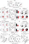
B6 or B6-Kd+/− WT mice were respectively transferred with 500 naïve OT-I-tomato+ and WP20-GFP+ T cells and immunized with WT Lm-Ova and Lm. Six wks later, purified OT-I or WP20 memory cells were adoptively transferred to CCR2-DTR mice. (A–C) Mice were further injected or not with clodronate liposomes and treated or not with DT prior to WT Lm or MCMV infection. At indicated times after the challenge infection, expression of IFN-γ and/or GrB by splenic OT-I and NK cells was monitored in the different experimental groups. Dot plot are representative FACS profiles after gating on OT-I or NK cells and numbers correspond to the % of IFN-γ+ or GrB+ cells in the gate shown. Scattered plots represent the pool of 2–3 independent replicate experiments (n=3–9) with p-values. In (D), WP20 memory cells were transferred to CCR2-DTR mice (+/−DT) expressing or not expressing the MHC-I molecule Kd prior to WT Lm infection. Grey dots depict resting memory CD8+ T cells. In (E), OT-I memory cells were transferred into CCR2-DTR mice (+/−DT) further challenged with WT ΔActA Lm-Ova and their frequency in the spleen of mice was measured 3 days later. Results represent 2–3 independent replicate experiments (n=2–5) with p-values.

Comment in

References

    1. Bajenoff M, Narni-Mancinelli E, Brau F, Lauvau G. Visualizing early splenic memory CD8+ T cells reactivation against intracellular bacteria in the mouse. PLoS One. 2010;5:e11524. - PMC - PubMed
    1. Barton ES, White DW, Cathelyn JS, Brett-McClellan KA, Engle M, Diamond MS, Miller VL, Virgin HWt. Herpesvirus latency confers symbiotic protection from bacterial infection. Nature. 2007;447:326–329. - PubMed
    1. Berg RE, Crossley E, Murray S, Forman J. Memory CD8+ T cells provide innate immune protection against Listeria monocytogenes in the absence of cognate antigen. J Exp Med. 2003;198:1583–1593. - PMC - PubMed
    1. Beuneu H, Deguine J, Bouvier I, Di Santo JP, Albert ML, Bousso P. A dual role for type I IFNs during polyinosinic-polycytidylic acid-induced NK cell activation. J Immunol. 2011;187:2084–2088. - PubMed
    1. Burkett PR, Koka R, Chien M, Chai S, Boone DL, Ma A. Coordinate expression and trans presentation of interleukin (IL)-15Ralpha and IL-15 supports natural killer cell and memory CD8+ T cell homeostasis. J Exp Med. 2004;200:825–834. - PMC - PubMed

Publication types

MeSH terms