. 2012 Nov 9;423(5):736-51.
doi: 10.1016/j.jmb.2012.08.020. Epub 2012 Aug 29.

The structure of human GALNS reveals the molecular basis for mucopolysaccharidosis IV A

Affiliations

The structure of human GALNS reveals the molecular basis for mucopolysaccharidosis IV A

Yadilette Rivera-Colón et al. J Mol Biol. .

Abstract

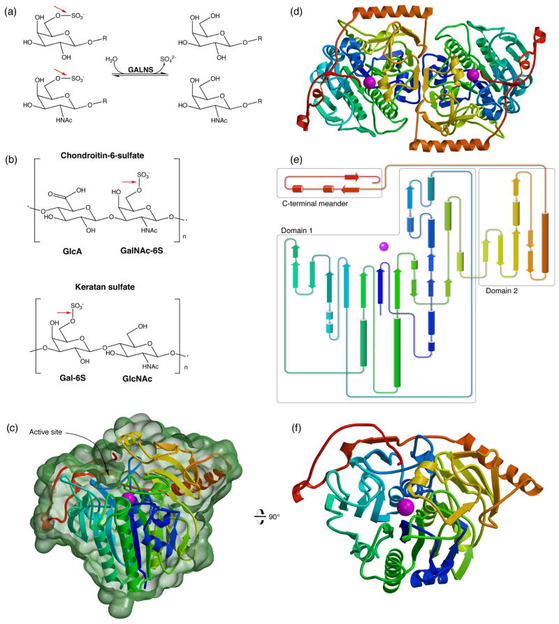
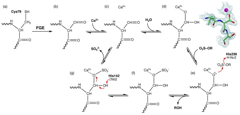
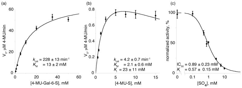
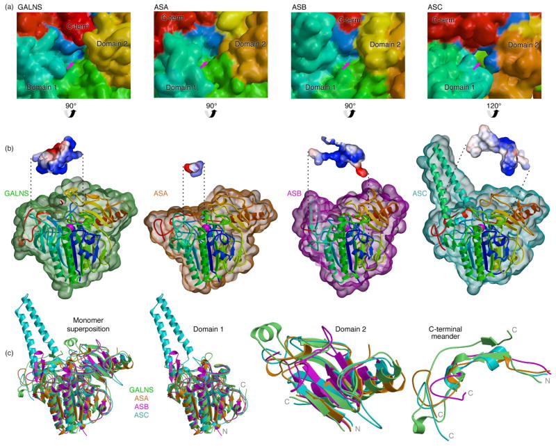
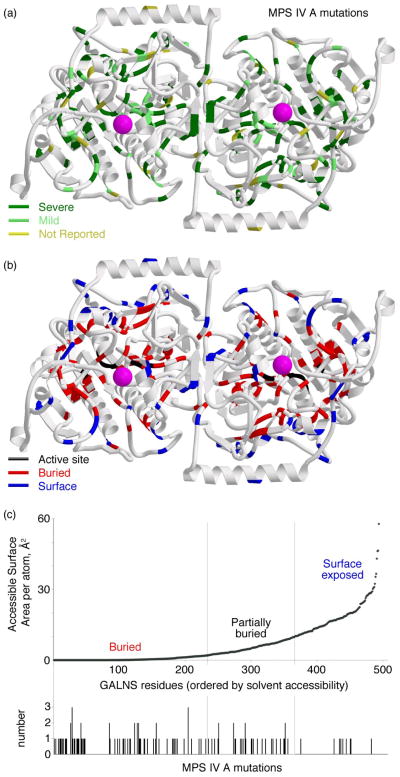
Lysosomal enzymes catalyze the breakdown of macromolecules in the cell. In humans, loss of activity of a lysosomal enzyme leads to an inherited metabolic defect known as a lysosomal storage disorder. The human lysosomal enzyme galactosamine-6-sulfatase (GALNS, also known as N-acetylgalactosamine-6-sulfatase and GalN6S; E.C. 3.1.6.4) is deficient in patients with the lysosomal storage disease mucopolysaccharidosis IV A (also known as MPS IV A and Morquio A). Here, we report the three-dimensional structure of human GALNS, determined by X-ray crystallography at 2.2Å resolution. The structure reveals a catalytic gem diol nucleophile derived from modification of a cysteine side chain. The active site of GALNS is a large, positively charged trench suitable for binding polyanionic substrates such as keratan sulfate and chondroitin-6-sulfate. Enzymatic assays on the insect-cell-expressed human GALNS indicate activity against synthetic substrates and inhibition by both substrate and product. Mapping 120 MPS IV A missense mutations onto the structure reveals that a majority of mutations affect the hydrophobic core of the structure, indicating that most MPS IV A cases result from misfolding of GALNS. Comparison of the structure of GALNS to paralogous sulfatases shows a wide variety of active-site geometries in the family but strict conservation of the catalytic machinery. Overall, the structure and the known mutations establish the molecular basis for MPS IV A and for the larger MPS family of diseases.


Figures

Fig. 1
Fig. 1
Human GALNS reaction and overall structure (a): GALNS cleaves 6-sulfate attached to Gal and GalNAc saccharides. (b): GALNS substrates include chondroitin-6-sulfate and keratan sulfate. Red arrows show the bond cleaved by GALNS. (c): The GALNS monomer structure colored from blue to red from N to C terminus and Ca2+ in magenta. (This coloring scheme is matched in subsequent panels.) The molecular surface in green (calculated in Povscript) is cut away to indicate the trench defining the active site. (d): An overview of the GALNS dimer structure viewed down the molecular dyad. (e): A topology diagram of the monomer shows domains boxed in grey. (f): An orthogonal view of the GALNS monomer viewed down the large β sheet in domain 1.
Fig. 2
Fig. 2
GALNS maturation and mechanism The GALNS gene encodes a cysteine at position 79 (a), which formylglycine generating enzyme (FGE) enzymatically converts into a formylglycine aldehyde (b). In the crystal, the aldehyde binds the calcium cofactor (c) and hydrates into the gem diol DHA (d). Substrate binding leads to nucleophilic attack on the sulfate (e), resulting of transfer of sulfate to the enzyme (f). Finally, intramolecular hydrolysis releases the sulfate (g) and regenerates the aldehyde. The protein in the crystal appears in state (d), while denaturing the enzyme for mass spectrometry disrupts the metal binding site and shifts the equilibrium to the aldehyde seen in state (b). The electron density in (d) is a σA-weighted 2Fo-Fc map contoured at 1.8s of the tripeptide centered at residue 79.
Fig. 3
Fig. 3
GALNS active site and ligand binding (a): Schematic of interactions in the GALNS active site. NCS-averaged interatomic distances in Å are shown in blue. (b) σA-weighted 2Fo-Fc map of the active site residues contoured at 1.8σ. (c): Ligand-bound structure. The electron density shows a σA-weighted 2Fo-Fc map contoured at 1.3σ around the GalNAc ligand. The molecular surface shows the large substrate-binding cavity with key residues indicated. (d): The GALNS electrostatic surface potential (calculated in Pymol) plotted from −57kT (red) to +57kT (blue) shows the large areas of positive charge suitable for interacting with polyanionic substrates. The active sites are indicated with arrows, and the inset shows the active site with GalNAc bound. A tetrasaccharide fragment of the keratan sulfate substrate (labeled “KS fragment”) is shown for scale.
Fig. 4
Fig. 4
GALNS reaction kinetics (a) and (b): Michaelis-Menten velocity vs. substrate concentration plots of GALNS with the synthetic substrates 4-MU-Gal-6-S (a) and 4-MU-S (b). 4-MU-S showed evidence of substrate inhibition (see text). (c): GALNS cleavage of 4-MU-S is inhibited by the presence of sulfate, which shows an IC50 of 0.89 mM in the assay.
Fig. 5
Fig. 5
Comparison of GALNS to other human sulfatases (a) Surface representations of GALNS (PDB ID 4FDI), ASA (PDB ID 1AUK), ASB (PDB ID 1FSU), and ASC (PDB ID 1P49), colored as in Fig. 1 and viewed into the active sites (indicated by arrows). (b) Surfaces of GALNS (green), ASA (orange), ASB (purple), and ASC (cyan) are shown around ribbon representation of the monomers (colored as in Fig. 1). The front surfaces are cropped to show the active site in the vicinity of the metal-binding site (magenta), and insets show the surface cavities (calculated in Pymol) colored by electrostatic potentials as in Fig. 3. (c) Superposition of GALNS (green), ASA (orange), ASB (purple), and ASC (cyan) are separated into panels corresponding to domain 1, domain 2, and the C-terminal meander, with the termini of each chain indicated.
Fig. 6
Fig. 6
GALNS mutations in MPS IV A (a): Disease-causing mutations leading to substitutions in the protein were mapped onto the GALNS structure. In the top panel, reported mutations are colored by their disease phenotype, as severe (less than 125cm in stature, dark green), mild (over 125cm, light green), or not reported (yellow). In the bottom panel, the mutations are mapped to their location on the protein, as near the active site (black), buried in the hydrophobic core (red), or on the surface (blue). (b): GALNS residues were ranked by solvent accessibility and vertical bars indicate a substitution leading to MPS IV A disease. Most of the mutations lead to changes in buried residues, meaning MPS IV A is most often a protein-folding disease.

References

    1. Neufeld EF, Muenzer J. The Mucopolysaccharidoses. In: Scriver CR, Beaudet AL, Sly WS, Valle D, editors. The Metabolic and Molecular Bases of Inherited Disease. 8. McGraw-Hill; New York: 2001. pp. 3421–3452.
    1. Northover H, Cowie RA, Wraith JE. Mucopolysaccharidosis type IVA (Morquio syndrome): a clinical review. J Inherit Metab Dis. 1996;19:357–65. - PubMed
    1. Tomatsu S, Montano AM, Nishioka T, Gutierrez MA, Pena OM, Tranda Firescu GG, Lopez P, Yamaguchi S, Noguchi A, Orii T. Mutation and polymorphism spectrum of the GALNS gene in mucopolysaccharidosis IVA (Morquio A) Hum Mutat. 2005;26:500–12. - PubMed
    1. Martell L, Lau K, Mei M, Burnett V, Decker C, Foehr ED. Biomarker analysis of Morquio syndrome: identification of disease state and drug responsive markers. Orphanet J Rare Dis. 2011;6:84. - PMC - PubMed
    1. Glössl J, Truppe W, Kresse H. Purification and properties of N-acetylgalactosamine 6-sulphate sulphatase from human placenta. Biochem J. 1979;181:37–46. - PMC - PubMed

Publication types