. 2012 Oct 10;20(10):1715-25.
doi: 10.1016/j.str.2012.07.016. Epub 2012 Aug 30.

Identification of unknown protein function using metabolite cocktail screening

Affiliations

Identification of unknown protein function using metabolite cocktail screening

Igor A Shumilin et al. Structure. .

Abstract

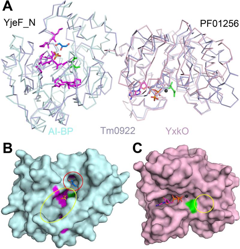
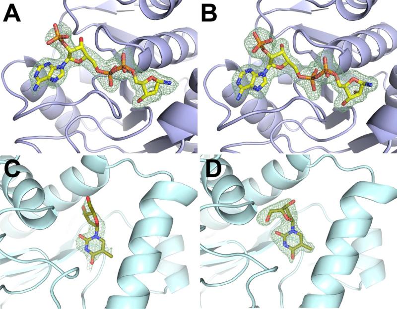
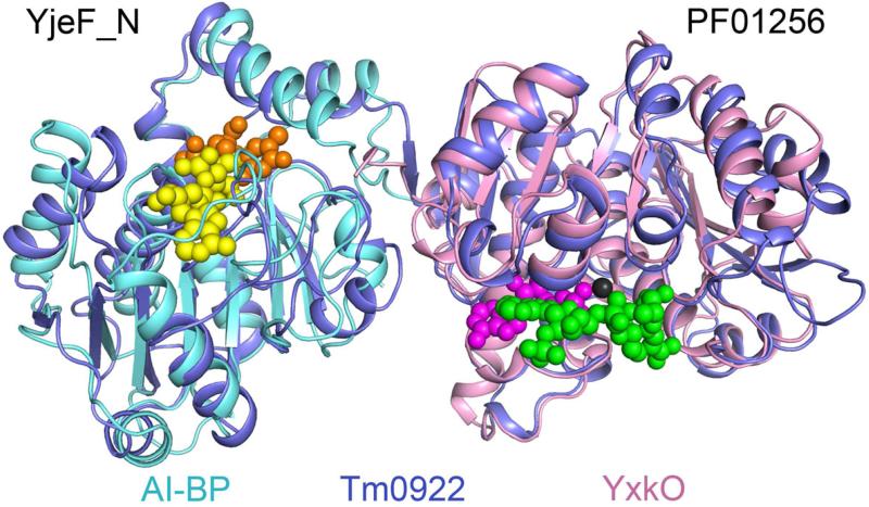
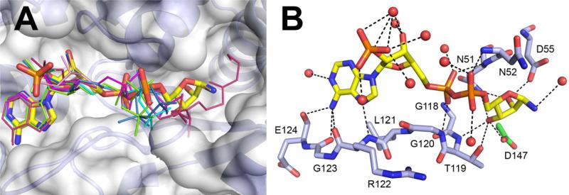
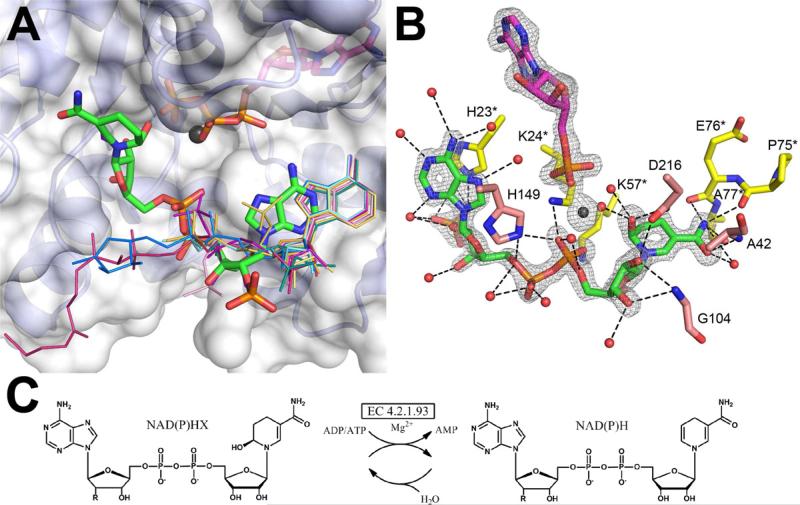
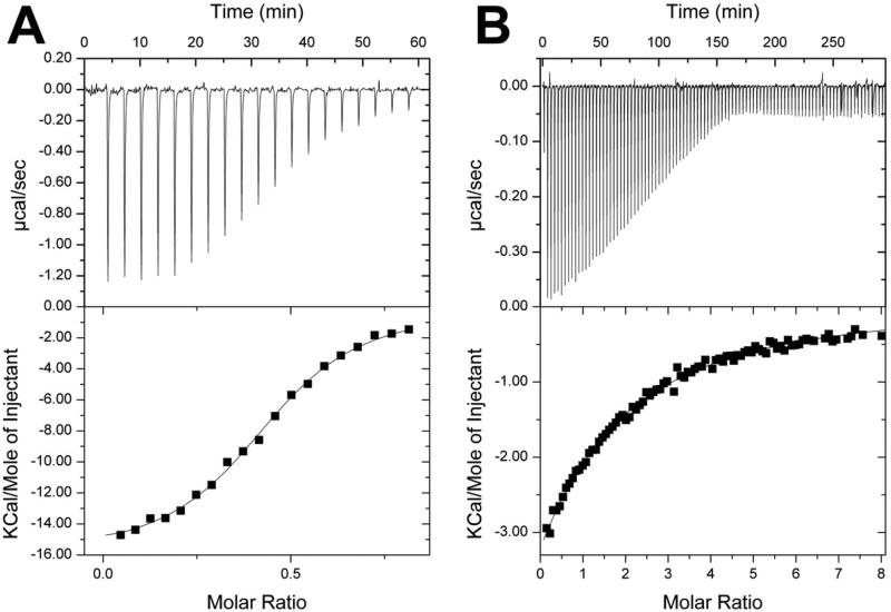
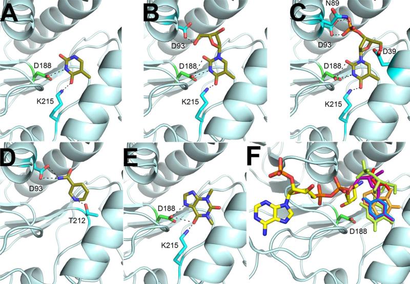
Proteins of unknown function comprise a significant fraction of sequenced genomes. Defining the roles of these proteins is vital to understanding cellular processes. Here, we describe a method to determine a protein function based on the identification of its natural ligand(s) by the crystallographic screening of the binding of a metabolite library, followed by a focused search in the metabolic space. The method was applied to two protein families with unknown function, PF01256 and YjeF_N. The PF01256 proteins, represented by YxkO from Bacillus subtilis and the C-terminal domain of Tm0922 from Thermotoga maritima, were shown to catalyze ADP/ATP-dependent NAD(P)H-hydrate dehydratation, a previously described orphan activity. The YjeF_N proteins, represented by mouse apolipoprotein A-I binding protein and the N-terminal domain of Tm0922, were found to interact with an adenosine diphosphoribose-related substrate and likely serve as ADP-ribosyltransferases. Crystallographic screening of metabolites serves as an efficient tool in functional analyses of uncharacterized proteins.


Figures

Figure 1
Figure 1
Subunit structures and putative active sites of the proteins from the YjeF_N and PF01256 families. (A) The superposition of Tm0922, a YjeF_N-PF01256 fusion (blue) with AI-BP, a single YjeF_N domain (cyan) and YxkO, a single PF01256 domain (pink). In the YjeF_N domain, the invariant D188AI-BP is green and highly conserved residues (Jha et al., 2008) are purple. The sulfate bound in the AI-BP structure is yellow/red and glycerol bound in the Tm0922 structure is navy/red. In the PF01256 domain, the invariant D216YxkO is green and the modeled ATP and Mg2+ (Zhang et al., 2002) are shown. (B) Surface of the AI-BP subunit colored as in Fig. 1A. The pocket compartment of the putative YjeF_N active site is marked by the red circle and its trench compartment is marked by the yellow oval. (C) Surface of the YxkO subunit colored as in Fig. 1A. The predicted kinase substrate binding site is marked by the yellow circle. The side chains of F162AI-BP and H149YxkO that block active sites in the apo-structures are removed.
Figure 2
Figure 2
Representative FO-FC omit electron density maps of the bound ligands in metabolite cocktail soaks and soaks with single ligands. (A) NADP is modeled in the NAD/NADP density in the Tm0922 crystal soaked with the cocktail G (resolution 2.05 Å, σ = 2.5). The disordered or hydrolyzed nicotinamide ring is not shown. (B) NADP soaked into the Tm0922 crystal (resolution 1.95 Å, σ = 2.5). (C). Thymidine is modeled in the thymine/thymidine density in the AI-BP crystal soaked with the cocktail E (resolution 2.5 Å, σ = 2.0). (D) Thymidine soaked into the AI-BP crystal (resolution 2.11 Å, σ = 2.5).
Figure 3
Figure 3
The binding sites of metabolitic ligands in the YjeF_N and PF01256 active sites. In the YjeF_N domain, ADP-ribose (yellow) marks the binding site for the “NAD set” of ligands while thymidine (orange) is bound at the “thymine set” site. In the PF01256 domain, bound AMP, Mg2+, and NADPHX are shown in magenta, black, and green, respectively.
Figure 4
Figure 4
Binding of the “NAD set” ligands in the YjeF_N site of Tm0922. (A) All ligands that contain an ADP moiety are bound in a similar mode along the trench compartment. NADP missing the nicotinamide ring is in a thick stick representation colored by the atom type. The other ligands (Table S1) are drawn as thin sticks and colored by the compound. Only the fragments visible in the electron density are shown. (B) The coordinating polar interactions of NADP. The invariant D147Tm0922 is green.
Figure 5
Figure 5
Binding of the “NAD set” metabolites in the PF01256 active sites and the reaction catalyzed by PF01256 proteins. (A) Ligands in the NAD(P)H soaks of Tm0922 bind in its kinase substrate site differently from the other ligands. NADPHX is in a thick stick representation colored by the atom type. The other ligands (Table S1) are drawn as thin sticks and colored by the compound. Ligands bound at the ATP binding site in the upper half are not shown except for ATP and Mg2+ from the ATP soak. Only the fragments visible in the electron density are shown. The second subunit of Tm0922 contributing to the PF01256 active site is not shown for clarity. (B) Coordination of NADPHX in the active site of YxkO, co-crystallized with ATP and Mg2+ and soaked with NADPH. The FO-FC omit electron density map calculated to 1.5 Å resolution and contoured at 4.0σ demonstrates the modification of the nicotinamide ring of NADPH and hydrolysis of ATP to AMP. Residues from two YxkO subunits forming the active site are colored in pink and yellow. (C) The reaction catalyzed by the orphan enzyme ATP-dependent NAD(P)H-hydrate dehydratase (EC 4.2.1.93) and spontaneous hydratation of NAD(P)H. R is a hydroxyl group in NADH/NADHX and a phosphate group in NADPH/NADPHX.
Figure 6
Figure 6
Isothermal titration calorimetry profiles and fitting curves for the binding of ADP-ribose to Tm0922 (A) and AI-BP (B) (Table 2).
Figure 7
Figure 7
Coordination of putative inhibitors in the YjeF_N site of AI-BP. Thymine (A), thymidine (B), thymidine 3′-monophosphate (C), nicotinamide (D) and theophylline (E) are colored by the atom type. (F) Binding of these ligands (colored by the compound) in the AI-BP active site would interfere with the binding of the NAD-related substrate due to the steric hindrance with a nicotinamide ring.

Comment in

References

    1. Acheson SA, Kirkman HN, Wolfenden R. Equilibrium of 5,6-hydration of NADH and mechanism of ATP-dependent dehydration. Biochemistry. 1988;27:7371–7375. - PubMed
    1. Anantharaman V, Aravind L. Novel conserved domains in proteins with predicted roles in eukaryotic cell-cycle regulation, decapping and RNA stability. BMC Genomics. 2004;5:45. - PMC - PubMed
    1. Carr R, Jhoti H. Structure-based screening of low-affinity compounds. Drug Discov Today. 2002;7:522–527. - PubMed
    1. Chance MR, Bresnick AR, Burley SK, Jiang JS, Lima CD, Sali A, Almo SC, Bonanno JB, Buglino JA, Boulton S, et al. Structural genomics: A pipeline for providing structures for the biologist. Protein Science. 2002;11:723–738. - PMC - PubMed
    1. Cheng G, Bennett EM, Begley TP, Ealick SE. Crystal structure of 4-amino-5-hydroxymethyl-2-methylpyrimidine phosphate kinase from Salmonella typhimurium at 2.3 A resolution. Structure. 2002;10:225–235. - PubMed

Publication types

MeSH terms

Associated data

LinkOut - more resources