. 2012 Oct 15;53(8):1584-94.
doi: 10.1016/j.freeradbiomed.2012.08.009. Epub 2012 Aug 23.

Nrf2 promotes alveolar mitochondrial biogenesis and resolution of lung injury in Staphylococcus aureus pneumonia in mice

Affiliations

Nrf2 promotes alveolar mitochondrial biogenesis and resolution of lung injury in Staphylococcus aureus pneumonia in mice

Janhavi Athale et al. Free Radic Biol Med. .

Abstract

Acute lung injury (ALI) initiates protective responses involving genes downstream of the Nrf2 (Nfe2l2) transcription factor, including heme oxygenase-1 (HO-1), which stimulates mitochondrial biogenesis and related anti-inflammatory processes. We examined mitochondrial biogenesis during Staphylococcus aureus pneumonia in mice and the effect of Nrf2 deficiency on lung mitochondrial biogenesis and resolution of lung inflammation. S. aureus pneumonia established by nasal insufflation of live bacteria was studied in mitochondrial reporter (mt-COX8-GFP) mice, wild-type (WT) mice, and Nrf2⁻/⁻ mice. Bronchoalveolar lavage, wet/dry ratios, real-time RT-PCR and Western analysis, immunohistochemistry, and fluorescence microscopy were performed on the lung at 0, 6, 24, and 48 h. The mice survived S. aureus inoculations at 5×10⁸ CFU despite diffuse lung inflammation and edema, but the Nrf2⁻/⁻ lung showed increased ALI. In mt-COX8-GFP mice, mitochondrial fluorescence was enhanced in bronchial and alveolar type II (AT2) epithelial cells. WT mice displayed rapid HO-1 upregulation and lower proinflammatory TNF-α, IL-1β, and CCL2 and, especially in AT2 cells, higher anti-inflammatory IL-10 and suppressor of cytokine signaling-3 than Nrf2⁻/⁻ mice. In the alveolar region, WT but not Nrf2⁻/⁻ mice showed strongly induced nuclear respiratory factor-1, PGC-1α, mitochondrial transcription factor-A, SOD2, Bnip3, mtDNA copy number, and citrate synthase. These findings indicate that S. aureus pneumonia induces Nrf2-dependent mitochondrial biogenesis in the alveolar region, mainly in AT2 cells. Absence of Nrf2 suppresses the alveolar transcriptional network for mitochondrial biogenesis and anti-inflammation, which worsens ALI. The findings link redox activation of mitochondrial biogenesis to ALI resolution.


Figures

Fig. 1
Fig. 1
Dose–response curve for ALI in WT mice as a function of S. aureus inoculation. (A) Bronchoalveolar lavage fluid (BALF) cell concentration (cells/ml × 105) plotted against S. aureus concentration. BALF cell concentration increased significantly at all levels of inoculation compared with control mice (*P<0.05). (B) Total BALF protein concentration plotted against S. aureus concentration. Protein concentrations increased significantly in mice inoculated with S. aureus at 5 × 10⁸ CFU (*P<0.05 vs control). Bars are means ± SEM.
Fig. 2
Fig. 2
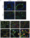
(A) Lung mitochondrial biogenesis in mt-COX8-GFP reporter mice after inoculation with 5 × 10⁸ CFU S. aureus (original magnification 40×). At 0 h, minimal green fluorescence is seen in airways and lung parenchyma by fluorescence microscopy. By 48 h, increased green fluorescence is detected in bronchial epithelium and in AT2 cells and macrophages. (B) Alveolar region of control lung shows GFP mitochondria, AT2 cell staining for SP-C (red), and nuclei (DAPI; blue) (image A); image B shows lung at 24 h after S. aureus inoculation; image C shows lung 24 h after breathing 250 ppm CO for 1 h as a positive control. Images D–F illustrate AT2 cells for conditions in images A–C (original magnification 100×).
Fig. 3
Fig. 3
Alveolar inflammation, acute lung injury, and edema in WT and Nrf2−/− mice after inoculation with 5 × 10⁸ CFU S. aureus. (A) BALF cell concentration (cells/ml × 105) is greater at baseline and increases more in Nrf2−/− than in WT mice. (B) Postinoculation BALF protein concentration increases significantly in both strains of mice (*P<0.05 vs control), but at 24 h, Nrf2−/− mice show a doubling of BALF protein compared with WT mice (**P<0.05 vs WT; bars are means ± SEM for n=4). (C) Lung wet-to-dry ratios in WT and Nrf2−/− mice increase significantly postinoculation (*P<0.05 vs control). At 6 h, Nrf2−/− mice demonstrate significantly more edema than WT mice (**P<0.05; values are means ± SEM for n=4 or 5 at each time point).
Fig. 4
Fig. 4
H&E-stained sections of inflation-fixed lung in WT and Nrf2−/− mice at 0 and 24 h postinoculation with 5 × 10⁸ CFU S. aureus (original magnification 20×). Compared with control, the lung’s inflammatory response is markedly enhanced in Nrf2−/− mice at 24 h (lower right). In that photomicrograph, loss of alveolar membrane integrity is indicated by red blood cell and plasma leakage (upper arrow) and alveolar neutrophil infiltration (lower arrow).
Fig. 5
Fig. 5
Comparison of lung tissue cytokine mRNA levels in WT and Nrf2−/− mice at 0 to 48 h after exposure to 5 × 10⁸ CFU S. aureus. Early phase lung cytokine levels increased at 6 to 24 h postinoculation followed at 48 h by full or partial resolution, except for CCL2 expression in Nrf2−/− mice. Compared with WT lung, the Nrf2−/− lung showed significantly greater expression of IL-1β, CCL2, and TNF-α and less expression of IL-10 (*P<0.05 compared with time 0, **P<0.05 vs WT control). Average fold change in expression is shown compared with lowest at time 0 (means ± SEM for n=4 samples each time point).
Fig. 6
Fig. 6
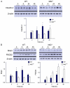
Pulmonary mitochondrial biogenesis after inoculation with 5 × 10⁸ CFU S. aureus. (A–E) Graphs of densitometry values for immunoblot analysis of lung parenchymal homogenates from WT and Nrf2−/− mice. (A) NRF-1 is rapidly induced in WT mice, but does not increase in Nrf2−/− mice until 48 h. (B) PGC-1α is rapidly induced in WT mice only. (C) Tfam is induced at 24 and 48 h in WT mice only. (D) WT lungs show early HO-1 induction, whereas Nrf2−/− lungs show late HO-1 induction. (E) SOD2 upregulation after S. aureus inoculation occurs in WT lungs, but not in Nrf2−/− lungs. All protein samples are normalized to β-actin. (F) Mitochondrial DNA copy number in the lung parenchyma after inoculation with 5 × 10⁸ CFU S. aureus. Copy number fails to increase by 48 h in the Nrf2−/− lung. For (A–F), bars are means ± SEM for n=4 per group per time point (*P<0.05 vs timed Nrf2−/− and the 0 time control; #P<0.05 vs the 0 time control only).
Fig. 7
Fig. 7
Alveolar claudin-4, mitochondrial autophagy, and programmed cell death responses by Western analysis of lung homogenates of WT and Nrf2−/− mice inoculated with 5 × 10⁸ CFU S. aureus. Protein samples are normalized to β-actin. (A) WT lungs display an early postinoculation increase in claudin-4, which is delayed until 48 h in Nrf2−/− lungs (graph of densitometry for n=4; *P<0.05 vs control and between strains). (B) S. aureus inoculation increases the mitochondrial autophagy protein Bnip3 in WT, but not in Nrf2−/− lungs. Concurrently lung PDCD2 protein increases in both mouse strains, but remains strongly elevated in Nrf2−/− mice for 48 h (graphs of densitometry for n=4; *P<0.05 vs control).
Fig. 8
Fig. 8
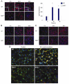
Fluorescence immunohistochemistry of WT and Nrf2−/− mouse lung sections pre- and postinoculation with 5 × 10⁸ CFU of S. aureus. Antibody-labeled protein appears in red with DAPI-stained nuclei in blue (original magnification 40×). (A) Citrate synthase (CS) staining of the mouse alveolar region. In WT lungs, CS is concentrated in cells with AT2 cell morphology (insets), especially at 24 and 48 h. In Nrf2−/− lungs, CS is detected mainly in mononuclear cells, most probably migrating macrophages. (B) IL-10 staining in lung tissue after S. aureus inoculation. In WT lungs, scattered IL-10 staining is found at 0 h, but increases at 24 and 48 h. IL-10 signal localizes mainly to alveolar macrophages at 24 h and AT2 cell staining is apparent by 48 h. Nrf2−/− lung demonstrates scattered alveolar IL-10 staining at baseline and minimal IL-10 induction after S. aureus. (C) SOCS3 staining in mouse alveolar region. SOCS3 is present constitutively in resident mononuclear cells in WT lung, but after S. aureus also appears in AT2 cells at 24 h and in type I and/or capillary epithelium at 48 h. SOCS3 is sparsely expressed by Nrf2−/− lungs, mainly in scattered cells of the alveolar region and, after S. aureus, increases in mononuclear inflammatory cells at 24 and 48 h. (D) SOCS3 immunochemical localization to AT2 cells (red) using surfactant protein C (SP-C, green). AT2 cells strongly express SOCS3 (yellow) at 24 h after inoculation of the lung with 5 × 10⁸ CFU of S. aureus.

References

    1. Kollef MH, Micek ST. Staphylococcus aureus pneumonia: a superbug infection in community and hospital settings. Chest. 2005;128:1093–1097. - PubMed
    1. Matthay MA, Zimmerman GA, Esmon C, Bhattacharya J, Coller B, Doerschuk CM, Floros J, Gimbrone MA, Hoffman E, Hubmayr RD, Leppert M, Matalon S, Munford R, Parsons P, Slutsky AS, Tracey KJ, Ward P, Gail DB, Harabin AL. Future research directions in acute lung injury: summary of a National Heart, Lung, and Blood Institute working group. Am. J. Respir. Crit. Care Med. 2003;167:1027–1035. - PubMed
    1. Matute-Bello G, Frevert CW, Martin TR. Animal models of acute lung injury. Am. J. Physiol. Lung Cell. Mol. Physiol. 2008;295:L379–399. - PMC - PubMed
    1. Demling RH. The modern version of adult respiratory distress syndrome. Annu. Rev. Med. 1995;46:193–202. - PubMed
    1. Fullerton JN, Singer M. Organ failure in the ICU: cellular alterations. Semin. Respir. Crit. Care Med. 2011;32:581–586. - PubMed

Publication types

MeSH terms