Review
. 2012 Oct-Dec;6(4):213-27.
doi: 10.4161/fly.21969. Epub 2012 Aug 31.

Drosophila egg chamber elongation: insights into how tissues and organs are shaped

Affiliations
Review

Drosophila egg chamber elongation: insights into how tissues and organs are shaped

Julie Gates. Fly (Austin). 2012 Oct-Dec.

Abstract

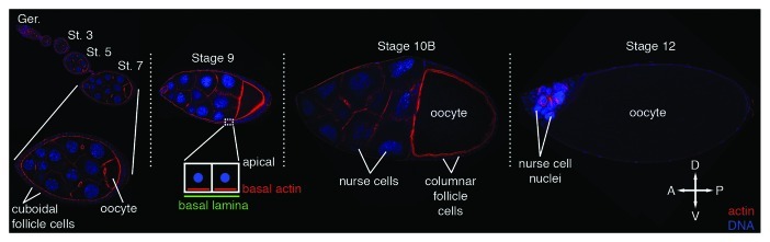
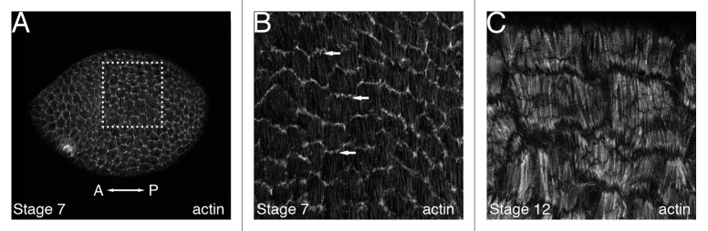
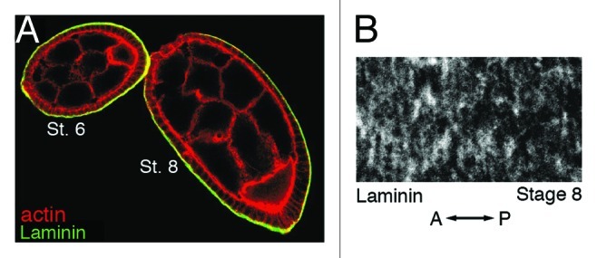
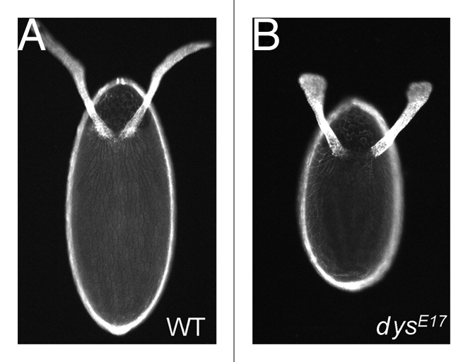
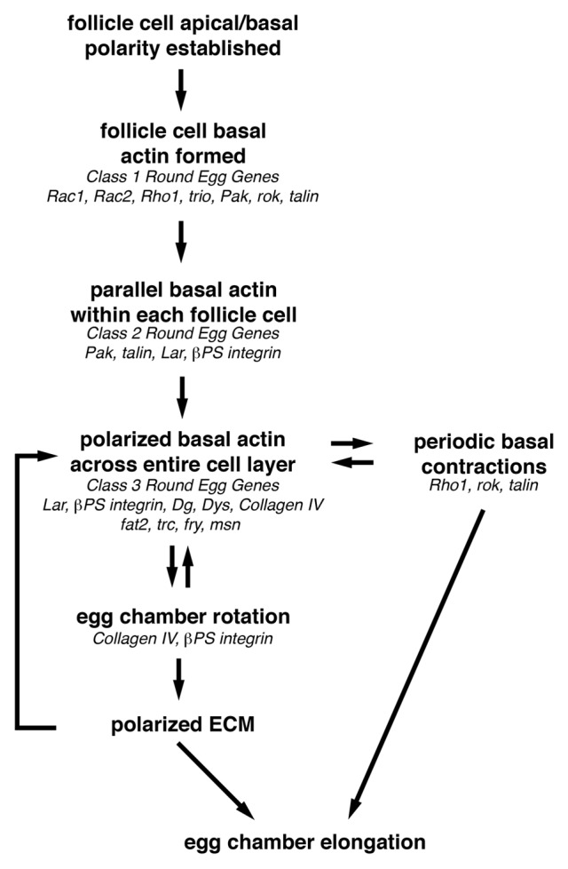
As tissues and organs are formed, they acquire a specific shape that plays an integral role in their ability to function properly. A relatively simple system that has been used to examine how tissues and organs are shaped is the formation of an elongated Drosophila egg. While it has been known for some time that Drosophila egg elongation requires interactions between a polarized intracellular basal actin network and a polarized extracellular network of basal lamina proteins, how these interactions contribute to egg elongation remained unclear. Recent studies using live imaging have revealed two novel processes, global tissue rotation and oscillating basal actomyosin contractions, which have provided significant insight into how the two polarized protein networks cooperate to produce an elongated egg. This review summarizes the proteins involved in Drosophila egg elongation and how this recent work has contributed to our current understanding of how egg elongation is achieved.


Figures

None
Figure 1. Drosophila oogenesis. Egg chambers from a subset of the stages of oogenesis, stained with phalloidin to visualize F-actin (red) and DAPI to visualize DNA (blue), reveal the major changes in the size and shape of the egg chamber and its component cells over time. A single focal plane through the center of each chamber is shown. Dotted lines separate egg chambers that were obtained from distinct ovarioles. A higher magnification image of the stage 7 egg chamber is included to show how the cuboidal follicle cell layer completely surrounds the germline cells at this stage. A similar organization of the egg chamber is seen from stages 1–8. The anterior/posterior (A/P) and dorsal/ventral (D/V) axes are indicated. Ger, germarium; St, stage.
None
Figure 2. Follicle cell basal actin filaments in wild-type egg chambers. The basal follicle cell surface of either an entire stage 7 egg chamber (A) or high magnification views of a subset of follicle cells from a stage 7 (B) or stage 12 (C) egg chamber that have been stained with phalloidin to visualize F-actin. All panels are oriented so that anterior is to the left. (B) High magnification view of the dotted box in (A) shows that the follicle cells in a stage 7 egg chamber produce actin-based protrusions (arrows) oriented in the same direction from a single-cell edge. These protrusions appear to reach out over the basal surface of the adjacent follicle cell. (C) High magnification view of the basal surface of a stage 12 egg chamber reveals basal actin filaments that are thicker and more pronounced than stage 7, with striations that resemble those seen in stress fibers in cultured cells.
None
Figure 3. Laminin is found along the outer basal surface of follicle cells and becomes oriented into strands that are perpendicular to the A/P axis of the egg chamber. (A) A stage 6 and stage 8 egg chamber that have been stained with phalloidin to visualize F-actin (red) and an antibody against the laminin α chain (green). (B) High magnification view of a stage 8 egg chamber that has been stained with an antibody against the laminin α chain. Orientation of the A/P axis of the egg chamber is indicated. St., Stage. (A and B) reprinted from reference with permission from Elsevier.
None
Figure 4. Round eggs are shorter and broader than wild-type. Dorsal view of eggs from wild-type (A) or dys mutant (dysE17/Df(3R)6184) (B) females. dys is a member of the class 3 round egg genes. Similar round eggs are produced when other round egg genes are mutated, irrespective of which class they belong to. Eggs are oriented with anterior at the top of the image. (A and B) reprinted from reference with permission from Elsevier.
None
Figure 5. Overview of how egg chamber elongation is achieved. Before egg chamber elongation can begin, a uniformly polarized network of follicle cell basal actin filaments must be established. This requires that the follicle cell basal domain be specified so that the basal actin filaments can be formed, then organized into parallel bundles within each follicle cell that then become oriented perpendicular to the A/P axis of the egg chamber. This uniformly polarized follicle cell basal actin network is then used to provide the force necessary to drive the active migration of the follicle cells over the stationary basal lamina during egg chamber rotation from stage 5–8. As the follicle cells migrate, they organize the ECM components of the basal lamina into parallel arrays that reinforce the orientation of the polarized basal actin filaments and form the molecular corset that contributes to egg chamber elongation. Then during stages 9 and 10, the polarized follicle cell basal actin filaments, together with non-muscle Myosin II, mediate the periodic basal contractions that may maintain the basal actin filaments and contribute to egg chamber elongation by augmenting the molecular corset around the center of the egg chamber. Genes that may function at each step of egg chamber elongation based on their loss-of-function phenotype are indicated in italics.
None
Figure 6. Loss of the round egg gene Lar results in follicle cell basal actin filaments that remain parallel within each cell but are no longer uniformly oriented across the follicle cell layer. High magnification views of a subset of follicle cells from a stage 12 wild-type egg chamber (A) or a stage 12 egg chamber that contains a clone of Lar mutant follicle cells (B). Both egg chambers have been stained with phalloidin to visualize F-actin (red) and an antibody against βPS integrin (green). Inset in (B) shows GFP marking wild-type cells at one-third scale. The mean orientation of actin filaments within wild-type cells in (B) is indicated with arrows. While some wild-type cells retain the proper orientation (far left cells), those closest to the mutant clone display defects in filament orientation, which resemble those in the Lar mutant cells (far right, without arrows). Orientation of the A/P axis of the egg chamber is indicated. (A and B) reprinted from reference with permission from Elsevier.
None
Figure 7. Model of egg chamber elongation. During oogenesis, egg chambers undergo a dramatic change in shape from a sphere (St. 4) to an ellipse (St. 10A). This is achieved through global rotation of the egg chamber within a static basal lamina (St. 5–8) and periodic contractions of a polarized basal actomyosin network (St. 9–10). Diagram depicts a cross section of egg chambers along with a high magnification view of the follicle cell basal surface for a subset of stages. Prior to the onset of egg chamber rotation, the follicle cell basal actin filaments (red) are parallel within each cell but are not uniformly oriented across the follicle cell layer and the ECM protein components of the follicle cell basal lamina (yellow) are unorganized. As the egg chamber rotates (green arrows indicate direction of rotation), the follicle cell basal actin filaments and the ECM fibers become aligned perpendicular to the A/P axis of the egg chamber forming the molecular corset. After the egg chamber stops rotating, a subset of follicle cells, concentrated around the widest point of the egg chamber, undergo periodic contractions of the polarized basal actin filaments, which are mediated by non-muscle Myosin II. These contractions result in a temporary change in the follicle cell basal surface and the generation of an inward force that reinforces the broader molecular corset. St., stage. Adapted from reference ,and reprinted with permission from Elsevier.

References

    1. Haigo SL, Bilder D. Global tissue revolutions in a morphogenetic movement controlling elongation. Science. 2011;331:1071–4. doi: 10.1126/science.1199424. - DOI - PMC - PubMed
    1. He L, Wang X, Tang HL, Montell DJ. Tissue elongation requires oscillating contractions of a basal actomyosin network. Nat Cell Biol. 2010;12:1133–42. doi: 10.1038/ncb2124. - DOI - PMC - PubMed
    1. Spradling A. Developmental Genetics of Oogenesis. In: Bate M, Martinez-Arias A, eds. The Development of Drosophila melanogaster Plainview, NY: Cold Spring Harbor Laboratory Press, 1993:1-70.
    1. Roth S, Lynch JA. Symmetry breaking during Drosophila oogenesis. Cold Spring Harb Perspect Biol. 2009;1:a001891. doi: 10.1101/cshperspect.a001891. - DOI - PMC - PubMed
    1. Mahajan-Miklos S, Cooley L. Intercellular cytoplasm transport during Drosophila oogenesis. Dev Biol. 1994;165:336–51. doi: 10.1006/dbio.1994.1257. - DOI - PubMed

LinkOut - more resources