. 2013 Feb;70(3):511-25.
doi: 10.1007/s00018-012-1141-z. Epub 2012 Sep 2.

EG-VEGF controls placental growth and survival in normal and pathological pregnancies: case of fetal growth restriction (FGR)

Affiliations

EG-VEGF controls placental growth and survival in normal and pathological pregnancies: case of fetal growth restriction (FGR)

S Brouillet et al. Cell Mol Life Sci. 2013 Feb.

Abstract

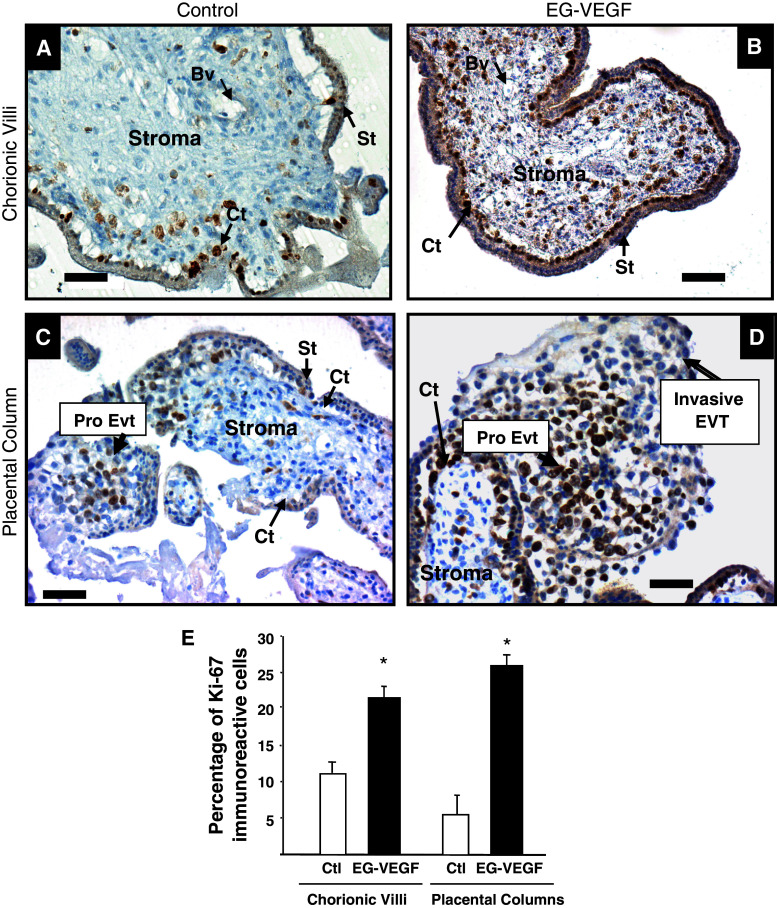
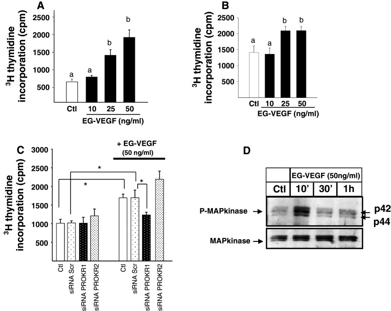
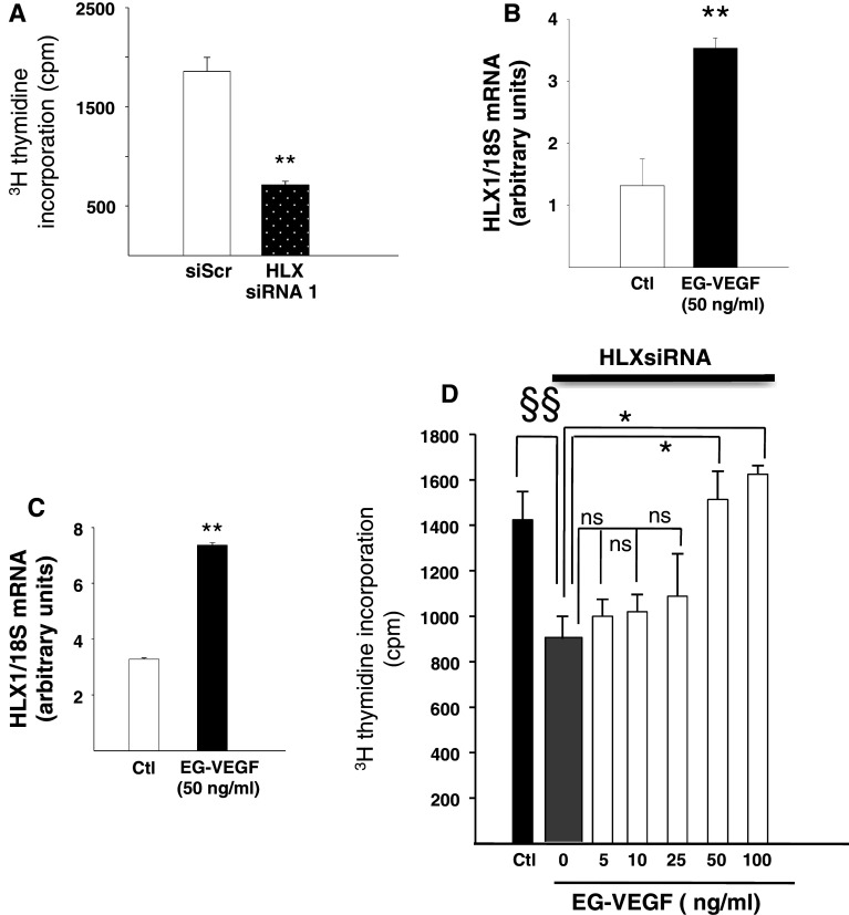
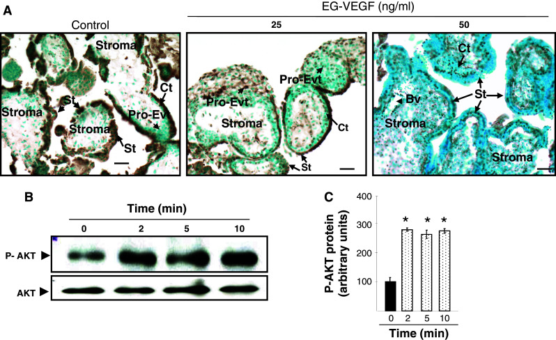
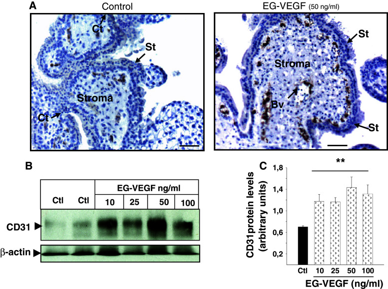
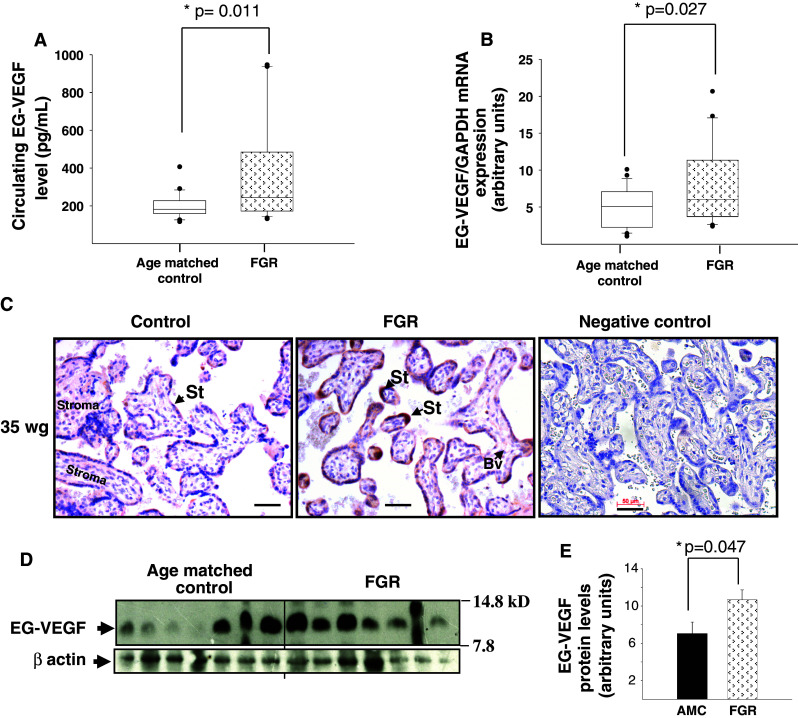
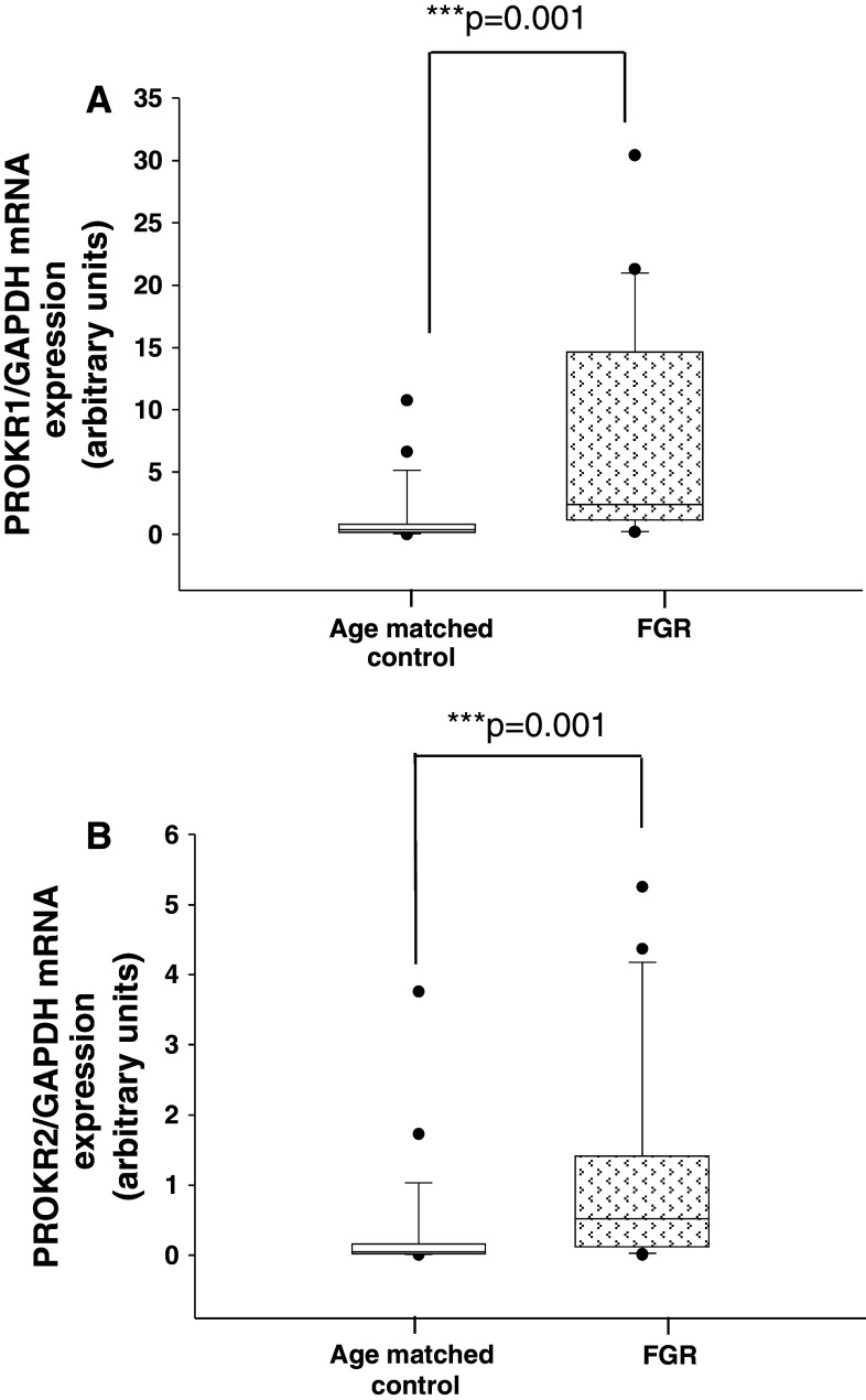
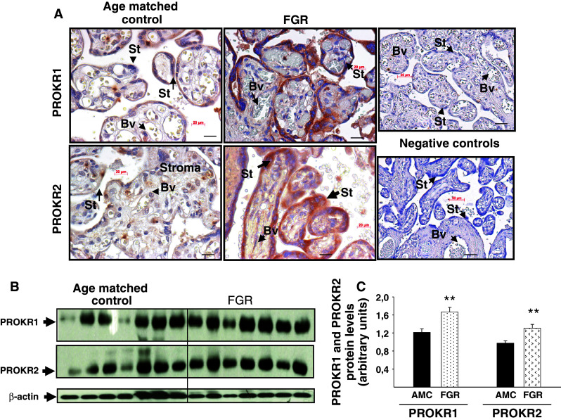
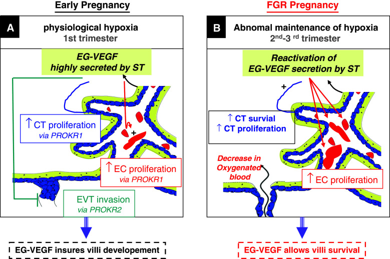
Identifiable causes of fetal growth restriction (FGR) account for 30 % of cases, but the remainders are idiopathic and are frequently associated with placental dysfunction. We have shown that the angiogenic factor endocrine gland-derived VEGF (EG-VEGF) and its receptors, prokineticin receptor 1 (PROKR1) and 2, (1) are abundantly expressed in human placenta, (2) are up-regulated by hypoxia, (3) control trophoblast invasion, and that EG-VEGF circulating levels are the highest during the first trimester of pregnancy, the period of important placental growth. These findings suggest that EG-VEGF/PROKR1 and 2 might be involved in normal and FGR placental development. To test this hypothesis, we used placental explants, primary trophoblast cultures, and placental and serum samples collected from FGR and age-matched control women. Our results show that (1) EG-VEGF increases trophoblast proliferation ([(3)H]-thymidine incorporation and Ki67-staining) via the homeobox-gene, HLX (2) the proliferative effect involves PROKR1 but not PROKR2, (3) EG-VEGF does not affect syncytium formation (measurement of syncytin 1 and 2 and β hCG production) (4) EG-VEGF increases the vascularization of the placental villi and insures their survival, (5) EG-VEGF, PROKR1, and PROKR2 mRNA and protein levels are significantly elevated in FGR placentas, and (6) EG-VEGF circulating levels are significantly higher in FGR patients. Altogether, our results identify EG-VEGF as a new placental growth factor acting during the first trimester of pregnancy, established its mechanism of action, and provide evidence for its deregulation in FGR. We propose that EG-VEGF/PROKR1 and 2 increases occur in FGR as a compensatory mechanism to insure proper pregnancy progress.


Figures

Fig. 1
Fig. 1
EG-VEGF increases the proliferation of villi and anchoring cytotrophoblast cells in PEX: the figure shows Ki-67 staining in PEX treated or not with 50 ng/ml EG-VEGF. a, c Ki67 staining in control placental villi and placental column, respectively. b, d The same staining under EG-VEGF treatment. e Percentage of Ki67-positive cytotrophoblast cells quantified in three independent experiments. Data represent the mean ± SEM of triplicates *p < 0.05 versus control. Scale bar: 50 μm. Ct cytotrophoblast, St syncytiotrophoblast, Bv blood vessel, Pro-Evt proliferative extravillous trophoblasts
Fig. 2
Fig. 2
EG-VEGF increases cytotrophoblast proliferation via PROKR1 but not PROKR2. a [3H]-Thymidine incorporation into cytotrophoblasts, in the absence (white bars) or presence of EG-VEGF at indicated concentrations (black bars) (*p < 0.05, **p < 0.01 vs. control). Data represent the mean ± SEM of triplicate determinations in three independent experiments. b Dose–response effect of EG-VEGF on BeWo cells proliferation. c [3H]-Thymidine incorporation into BeWo cells that have been transfected with scramble or siRNA for PROKR1 or PROKR2 and treated with EG-VEGF (*p < 0.05). Data represent the mean ± SEM. d Representative Western blot of MAP kinase phosphorylations after treatment with 50 ng/ml of EG-VEGF of cytotrophoblasts. Standardization of the protein signals was done with antibodies against total-MAP kinases. Data are expressed as mean + SE. Values overwritten with different letters are significantly different from each other (p < 0.05)
Fig. 3
Fig. 3
EG-VEGF effect on HLX expression in PEX and in BeWos. a Effect of HLX inactivation on BeWo proliferation. b, c Effect of EG-VEGF on HLX mRNA expression in PEX cells and in BeWo, respectively. d Depiction of EG-VEGF concentration-dependent (5–100 ng/ml) rescue of proliferation of BeWo cells in the presence of HLX inactivation using siRNA. Data are expressed as mean ± SEM. §§Corresponds to the comparison between the control and the HLXsiRNA in the absence of EG-VEGF. *Represents the comparison between the HLXsiRNA in the absence and in the presence of EG-VEGF (50 and 100 ng/ml). ns Corresponds to the comparison between the HLXsiRNA in the absence and in the presence of EG-VEGF (5–25 ng/ml) (*p < 0.05, **p < 0.01 vs. control)
Fig. 4
Fig. 4
EG-VEGF effect on placental villi survival. a Representative photographs of TUNEL staining (in brown) in PEX that have been treated or not with EG-VEGF. PEX (10 wg) were serum starved for 72 h and treated for 24 h with EG-VEGF (25 and 50 ng/ml). EG-VEGF condition shows much less TUNEL-positive cells compared to the control condition. b Representative Western blot of AKT phosphorylation after treatment with 50 ng/ml EG-VEGF. Standardization of the protein signals was done with antibodies against total-AKT. Quantification of the intensity of the bands is illustrated in c. Data represent the mean ± SEM of triplicates *p < 0.05 vs. control. Scale bar: 20 μm. Ct cytotrophoblast, St syncytiotrophoblast, Bv blood vessel, Pro-Evt proliferative extravillous trophoblasts
Fig. 5
Fig. 5
EG-VEGF effect on the vascularization of first-trimester placental villi. a Representative photographs of CD31 staining (in brown) in PEX that have been treated or not with EG-VEGF. b Representative Western blot of CD31 expression after treatment with 10–50 and 100 ng/ml of EG-VEGF. Standardization of the protein signals was done with antibodies against β actin. Quantification of the intensity of the bands is illustrated in c. Data represent the mean ± SEM of triplicates *p < 0.05 vs. control. Scale bar: 50 μm. Ct cytotrophoblast, St syncytiotrophoblast, Bv blood vessel
Fig. 6
Fig. 6
EG-VEGF expression is up-regulated in FGR pregnancies. a EG-VEGF serum levels in FGR and AMC pregnant women. EG-VEGF levels were measured by ELISA (*p < 0.05). b EG-VEGF mRNA levels in FGR and AMC placentas (25 AMC and 21 FGR) (*p < 0.05). c Chorionic villi sections immunostained with anti-EG-VEGF antibody. d Representative Western-blot analysis that compares AMC versus FGR placentas (14 AMC and 14 FGR). Quantification of the intensity of the bands is illustrated in e (*p < 0.05). Villous cytotrophoblasts and syncytiotrophoblasts were positively stained. Scale bar: 50 μm. Ct cytotrophoblast, St syncytiotrophoblast, Bv blood vessel
Fig. 7
Fig. 7
PROKR1 and PROKR2 mRNA expression in placental tissue from AMC and FGR pregnancies. a Quantification of PROKR1 mRNA levels expression in placental tissues from AMC and FGR pregnancies. b Quantification of PROKR2 mRNA levels expression in placental tissues from AMC and FGR pregnancies in the third trimester. We analyzed 25 AMC and 21 FGR placentas
Fig. 8
Fig. 8
PROKR1 and PROKR2 protein expression in placental tissue from AMC and FGR pregnancies. a Representative photographs of PROKR1 and PROKR2 staining in AMC and FGR placentas. b Representative Western blots of PROKR1 and PROKR2 expression in AMC and FGR (14 AMC and 14 FGR). Standardization of the protein signals was done using beta actin. Quantification of the intensity of the bands is illustrated in c. Data represent the mean ± SEM of triplicate *p < 0.05 versus control. Ct cytotrophoblast, St syncytiotrophoblast, Bv blood vessel
Fig. 9
Fig. 9
A proposed model for EG-VEGF-mediated effects in human placenta during the first trimester of pregnancy and in FGR. During the first trimester of human pregnancy (a), EG-VEGF is highly secreted from the ST layer and stimulates cytotrophoblast and endothelial cell proliferation via PROKR1, respectively. EG-VEGF also inhibits EVT invasion. These local effects contribute to the growth of the villi and to the formation of trophoblast plugs within the maternal spiral arteries; this protects the villi from the potentially harmful effects of oxygen species against which the placenta is not yet protected. In FGR placentas (b), we showed that EG-VEGF and its receptors are up-regulated, we propose that these increases occur as responses to hypoxia associated with this pathology, and that EG-VEGF and its receptors play a compensatory role to allow villi growth through effects on CT and endothelial cells proliferation, a process strongly affected in FGR. The EG-VEGF/PROKRs system acts then as survival machinery, central to the pregnancy progress

References

    1. Cross JC, Baczyk D, Dobric N, Hemberger M, Hughes M, Simmons DG, Yamamoto H, Kingdom JC. Genes, development and evolution of the placenta. Placenta. 2003;24(2–3):123–130. - PubMed
    1. Henriksen T. Foetal nutrition, foetal growth restriction and health later in life. Acta Paediatr Suppl. 1999;88(429):4–8. - PubMed
    1. Barker DJ. Fetal growth and adult disease. Br J Obstet Gynaecol. 1992;99(4):275–276. - PubMed
    1. Bernstein IM, Horbar JD, Badger GJ, Ohlsson A, Golan A. Morbidity and mortality among very-low-birth-weight neonates with intrauterine growth restriction. The Vermont Oxford Network. Am J Obstet Gynecol. 2000;182(1 Pt 1):198–206. - PubMed
    1. Baschat AA. Fetal responses to placental insufficiency: an update. BJOG. 2004;111(10):1031–1041. - PubMed

Publication types

MeSH terms