. 2012 Oct;8(10):855-61.
doi: 10.1038/nchembio.1062.

A new type V toxin-antitoxin system where mRNA for toxin GhoT is cleaved by antitoxin GhoS

Affiliations

A new type V toxin-antitoxin system where mRNA for toxin GhoT is cleaved by antitoxin GhoS

Xiaoxue Wang et al. Nat Chem Biol. 2012 Oct.

Abstract

Among bacterial toxin-antitoxin systems, to date no antitoxin has been identified that functions by cleaving toxin mRNA. Here we show that YjdO (renamed GhoT) is a membrane lytic peptide that causes ghost cell formation (lysed cells with damaged membranes) and increases persistence (persister cells are tolerant to antibiotics without undergoing genetic change). GhoT is part of a new toxin-antitoxin system with YjdK (renamed GhoS) because in vitro RNA degradation studies, quantitative real-time reverse-transcription PCR and whole-transcriptome studies revealed that GhoS masks GhoT toxicity by cleaving specifically yjdO (ghoT) mRNA. Alanine substitutions showed that Arg28 is important for GhoS activity, and RNA sequencing indicated that the GhoS cleavage site is rich in U and A. The NMR structure of GhoS indicates it is related to the CRISPR-associated-2 RNase, and GhoS is a monomer. Hence, GhoT-GhoS is to our knowledge the first type V toxin-antitoxin system where a protein antitoxin inhibits the toxin by cleaving specifically its mRNA.


Figures

Fig. 1
Fig. 1. GhoT increases persistence
Cell survival (%) after ampicillin (100 µg/mL) treatment for 2, 4, and 6 h with MqsR production with and without ghoT (a), or with GhoT production (b). wt indicates the wild-type host (E. coli BW25113). (c) Revival of GhoT-induced persister cells was tested by producing GhoT in BW25113 ΔghoT/pCA24N-ghoT while treating cells with ampicillin (100 µg/ml) for 2 hours. Growth in fresh LB medium was compared to control cells that lacked ampicillin treatment. At least three independent cultures of each strain were evaluated for each experiment, and error bars indicate standard error of mean.
Fig. 2
Fig. 2. GhoT is toxic and GhoS reduces this toxicity
(a) Cell growth in LB medium for cells producing GhoT and GhoS. Note the chromosomal copy of ghoS in the wild-type strain allows for some growth with toxin GhoT production. GhoSX is truncated GhoS with a stop codon introduced at Tyr16. Three independent cultures of each strain were evaluated, and error bars indicate standard error of mean (n = 3). (b) Cell culture at the end of growth in (a) at 20 h to show the clearance and lysis due to production of GhoT. Scale bar represents 1 cm. (c) Cell morphology after incubating for 8 h at 37°C. Scale bar represents 5 µm. For (a), (b) and (c), Empty: BW25113/pCA24N/pBS(Kan), GhoT: BW25113/pCA24N-ghoT/pBS(Kan), GhoS: BW25113/pCA24N/pBS(Kan)-ghoS, GhoT + GhoS: BW25113/pCA24N-ghoT/pBS(Kan)-ghoS, and GhoT + GhoSX: BW25113/pCA24N-ghoT/pBS(Kan)-ghoSX. Plasmids were retained with kanamycin (50 µg/mL) and chloramphenicol (30 µg/mL); 0.5 mM IPTG was used at time 0 to produce the plasmid-based proteins. Three independent cultures of each strain were evaluated. (d) Growth on LB plates with kanamycin (50 µg/mL), chloramphenicol (30 µg/mL), and IPTG (1 mM, to induce ghoT via pCA24N-ghoT). In the absence of a chromosomal copy of ghoS, there is no growth with toxin GhoT production. ΔghoS is BW25113 ΔghoS and ΔghoT is BW25113 ΔghoT. p refers to pCA24N and p-ghoT refers to pCA24N-ghoT, respectively. Three independent cultures of each strain were evaluated. Scale bar represents 1 cm.
Fig. 3
Fig. 3. GhoS adopts a ferredoxin-like fold and Arg28 is important for its cleavage activity
(a) Ribbon model of the lowest-energy conformer of GhoS, with the secondary structural elements and termini labeled; putative catalytically important residues shown as sticks and labeled. Figure prepared with PyMOL (http://www.pymol.org/). (b) Two-micrograms of in vitro synthesized wild-type ghoT transcript (207 nt, lane 1) were incubated without (−) or with 30 µg of purified GhoS and its variants at 37°C for 3 h. Two mutants, F14A and F55A, eluted from the size exclusion column as monomers (M) and dimers (D), so both forms were tested (F14A, 40% dimer; F55A, 32% dimer). The reduced activity of GhoS with point mutations is shown by the presence of un-cleaved transcript as indicated by an arrow. M indicates low range ssRNA ladder. (c) Circular dichroism (CD) spectra demonstrating that native GhoS (dark blue) and all the GhoS mutants are folded (sample concentrations ~20 µM). (d) Co-expression of GhoT with wild-type (WT) GhoS and the GhoS variants via BL21(DE3)/pCA24N-ghoT harboring the pRP1B(Kan)-ghoS constructs (0.1 mM IPTG was used). Scale bar represents 1.1 cm.
Fig. 4
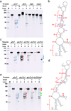
Fig. 4. GhoS cleavage of native and altered ghoT transcripts
(a) GhoS cleavage reaction with native transcripts of ghoT (207 nt), ghoS (311 nt), atpE (189 nt) and ompA (211 nt). HI indicates heat inactivated GhoS. The blue arrows indicate the main fragments generated after cleavage. M indicates the low range ssRNA ladder. (b) Predicted secondary structure of in vitro synthesized ghoT mRNA. Capital red letters indicate the changed nt for mutations m1, m2, m3, and m4. S1, S2, S3, S4, and S5 indicate the cleavage sites based on RNA sequencing. The four main sections in the structure are indicated with numbers i, ii, iii and iv. The RNA secondary structure was obtained using Mfold software. (c) GhoS cleavage reaction with transcripts of ghoT with mutations m1, m2, m3, and m4 (207 nt). The red arrows indicate the fragments generated or increased in the mutant transcripts after cleavage. (d) Predicted secondary structure of in vitro synthesized ghoTm1m2 mRNA. The mutated ghoTm1m2 cleavage site is indicated by two solid red lines. (e) GhoS cleavage reaction with transcripts of ghoT with mutations m1, m2, and m1m2 (207 nt). The green arrows indicate the reduced fragments after the introduction of the second mutation. For the reactions shown in (a), (c), and (e), 2 µg of in vitro synthesized transcripts were incubated in with (−) or without 30 µg (+) of purified GhoS at 37°C for 3 h and analyzed by gel electrophoresis.

Comment in

  • GhoSTly bacterial persisters.
    Van Melderen L. Van Melderen L. Nat Chem Biol. 2012 Oct;8(10):812-3. doi: 10.1038/nchembio.1066. Nat Chem Biol. 2012. PMID: 22987009

References

    1. Gerdes K, Christensen SK, Lobner-Olesen A. Prokaryotic toxin-antitoxin stress response loci. Nat. Rev. Microbiol. 2005;3:371–382. - PubMed
    1. Hayes F, Van Melderen L. Toxins-antitoxins: diversity, evolution and function. Crit. Rev. Biochem. Mol. Biol. 2011;46:386–408. - PubMed
    1. Masuda H, Tan Q, Awano N, Wu K-P, Inouye M. YeeU enhances the bundling of cytoskeletal polymers of MreB and FtsZ, antagonizing the CbtA (YeeV) toxicity in Escherichia coli. Mol. Microbiol. 2012;84:979–989. - PubMed
    1. Ren D, Bedzyk LA, Thomas SM, Ye RW, Wood TK. Gene expression in Escherichia coli biofilms. Appl. Microbiol. Biotechnol. 2004;64:515–524. - PubMed
    1. Kim Y, Wang X, Qun M, Zhang X-S, Wood TK. Toxin-antitoxin systems in Escherichia coli influence biofilm formation through YjgK (TabA) and fimbriae. J. Bacteriol. 2008;191:1258–1267. - PMC - PubMed

Publication types

Associated data