. 2012 Nov;78(21):7760-8.
doi: 10.1128/AEM.01858-12. Epub 2012 Aug 31.

OmpA-mediated biofilm formation is essential for the commensal bacterium Sodalis glossinidius to colonize the tsetse fly gut

Affiliations

OmpA-mediated biofilm formation is essential for the commensal bacterium Sodalis glossinidius to colonize the tsetse fly gut

Michele A Maltz et al. Appl Environ Microbiol. 2012 Nov.

Abstract

Many bacteria successfully colonize animals by forming protective biofilms. Molecular processes that underlie the formation and function of biofilms in pathogenic bacteria are well characterized. In contrast, the relationship between biofilms and host colonization by symbiotic bacteria is less well understood. Tsetse flies (Glossina spp.) house 3 maternally transmitted symbionts, one of which is a commensal (Sodalis glossinidius) found in several host tissues, including the gut. We determined that Sodalis forms biofilms in the tsetse gut and that this process is influenced by the Sodalis outer membrane protein A (OmpA). Mutant Sodalis strains that do not produce OmpA (Sodalis ΔOmpA mutants) fail to form biofilms in vitro and are unable to colonize the tsetse gut unless endogenous symbiotic bacteria are present. Our data indicate that in the absence of biofilms, Sodalis ΔOmpA mutant cells are exposed to and eliminated by tsetse's innate immune system, suggesting that biofilms help Sodalis evade the host immune system. Tsetse is the sole vector of pathogenic African trypanosomes, which also reside in the fly gut. Acquiring a better understanding of the dynamics that promote Sodalis colonization of the tsetse gut may enhance the development of novel disease control strategies.


Figures

Fig 1
Fig 1
In situ visualization of Sodalis clusters present in the tsetse gut. (A) Cross sections of tsetse gut tissue stained with DAPI. DNA from host epithelial tissue and Sodalis cells all stains blue. Bar, 50 μm. (B) Visualization of Sodalis clusters in the lumen of the tsetse gut using fluorescent in situ hybridization. Tissues were probed with a rhodamine-labeled oligonucleotide specific for Sodalis 16S rRNA (red). Bar, 50 μm. Autofluorescent blood meal-derived erythrocytes surround Sodalis cells. Bars, 50 μm. (C) Panels A and B merged to better orient the position of Sodalis cell clusters in relation to tsetse gut epithelial tissues. In each of panels A, B, and C, the most prominent cluster of Sodalis cells is indicated by a white arrow. (D to F) Enlarged views of the Sodalis clusters shown in panels A to C, respectively. Bars, 10 μm.
Fig 2
Fig 2
Lectin staining of a biofilm-associated polysaccharide matrix surrounding a cluster of Sodalis cells. (A) A cross section of the tsetse gut tissue stained with a rhodamine-labeled oligonucleotide specific for Sodalis 16S rRNA. (B) A WGA lectin probe (green) bound to an extracellular polysaccharide matrix. (C) Panels A and B merged. The presence of a polysaccharide matrix surrounding a Sodalis cluster in the lumen of the tsetse gut indicates that these bacterial cells have formed a biofilm. Bars, 50 μm.
Fig 3
Fig 3
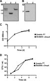
Generation of a Sodalis ompA mutant strain. (A) PCR amplification of the ompA gene in the Sodalis parent strain (Sodalis PT; lane 2) and Sodalis ompA mutants (Sodalis ΔOmpA; lane 3). The ∼1-kb band shift is the result of intron insertion into the ompA locus of the Sodalis chromosome. (B) A Western blot showing OmpA expression in Sodalis PT cells (lane 1) but not in their Sodalis ΔOmpA derivatives (lane 2). (C and D) Sodalis PT and Sodalis ΔOmpA growth curves in MM medium and blood, respectively. Sodalis ΔOmpA does not exhibit a growth defect when cultured in either medium.
Fig 4
Fig 4
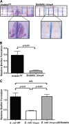
OmpA regulates bacterial biofilm formation in vitro. (A) Sodalis PT and Sodalis ΔOmpA cells were shaken vigorously at an angle for 7 days in 15-ml Falcon tubes. The bacterial splash zone was subsequently stained with crystal violet to visualize the presence or absence of a biofilm. These structures were present in the tubes containing Sodalis PT cells (shown enlarged in the corresponding box). Sodalis ΔOmpA cells failed to form a biofilm. This experiment was performed in triplicate with 3 distinct clonal populations of each bacterial strain. (B) Spectrophotometric quantification of relative biofilm formation exhibited by Sodalis PT and Sodalis ΔOmpA cells. (C) E. coli ΔOmpA cells exhibit a biofilm formation defect that is restored in their counterparts that were genetically modified to express Sodalis ompA (E. coli ΔOmpA pIOSodalis cells). In panels B, C and D, statistical significance was determined by the Mann-Whitney test.
Fig 5
Fig 5
OmpA modulates Sodalis biofilm formation in the tsetse gut. (A) A newly developed per os colonization assay was used to determine if Sodalis PT cells could establish an infection in GmmWT and GmmApo fly guts. Five days postinoculation, Sodalis PT was found at a density of ∼106 cells in both host backgrounds. (B) Biofilm formation in the tsetse gut was visualized by orally inoculating GmmWT and GmmApo flies with a strain of Sodalis (Sodalis pGFP) that expresses green fluorescent protein. Fluorescing microcolonies, indicated by a white arrow, were present in GmmWT flies 1 day postinoculation with Sodalis pGFP. Three days postinoculation, these microcolonies are more pronounced in GmmWT flies. Additionally, Sodalis pGFP colonies are also beginning to form at this time in GmmApo flies. Bars, 50 μm. (C) Colonization of GmmWT fly guts by Sodalis PT and Sodalis ΔOmpA strains. Sodalis ΔOmpA cells exhibit an initial colonization defect but, by 5 days postinoculation, reach a density similar to that of Sodalis PT cells. (D) Colonization of GmmSgm fly guts by Sodalis PT and Sodalis ΔOmpA strains. Sodalis ΔOmpA cells cannot colonize tsetse when their native counterparts are absent. In panel A, colonization assays were performed in duplicate and data are presented as the means ± standard deviations. In panels C and D, each symbol represents one fly gut, and the dotted line indicates the assay's limit of detection. Statistical significance in C and D was determined by the Mann-Whitney test.
Fig 6
Fig 6
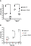
Biofilms protect Sodalis from tsetse's innate immune response. Sodalis PT and Sodalis ΔOmpA cell colonization of immune-activated GmmWT fly guts. LPS and PGN (A) or live E. coli (B) was fed to flies prior to bacterial inoculation. Guts were dissected at 1 and 3 days postinoculation to determine the number of CFU present. Each symbol represents one fly gut, and the dotted line indicates the assay's limit of detection. Statistical significance was determined by the Mann-Whitney test.

References

    1. Akman L, Rio RV, Beard CB, Aksoy S. 2001. Genome size determination and coding capacity of Sodalis glossinidius, an enteric symbiont of tsetse flies, as revealed by hybridization to Escherichia coli gene arrays. J. Bacteriol. 183:4517–4525 - PMC - PubMed
    1. Akman L, et al. 2002. Genome sequence of the endocellular obligate symbiont of tsetse flies, Wigglesworthia glossinidia. Nat. Genet. 32:402–407 - PubMed
    1. Aksoy S, Pourhosseini AA, Chow A. 1995. Mycetome endosymbionts of tsetse flies constitute a distinct lineage related to Enterobacteriaceae. Insect Mol. Biol. 4:15–22 - PubMed
    1. Attardo GM, et al. 2008. Analysis of milk gland structure and function in Glossina morsitans: milk protein production, symbiont populations and fecundity. J. Insect Physiol. 54:1236–1242 - PMC - PubMed
    1. Barrios AF, Zuo R, Ren D, Wood TK. 2006. Hha, YbaJ, and OmpA regulate Escherichia coli K12 biofilm formation and conjugation plasmids abolish motility. Biotechnol. Bioeng. 93:188–200 - PubMed

Publication types

Substances

LinkOut - more resources