. 2012 Oct;15(10):1439-44.
doi: 10.1038/nn.3203. Epub 2012 Sep 2.

Biasing the content of hippocampal replay during sleep

Affiliations

Biasing the content of hippocampal replay during sleep

Daniel Bendor et al. Nat Neurosci. 2012 Oct.

Abstract

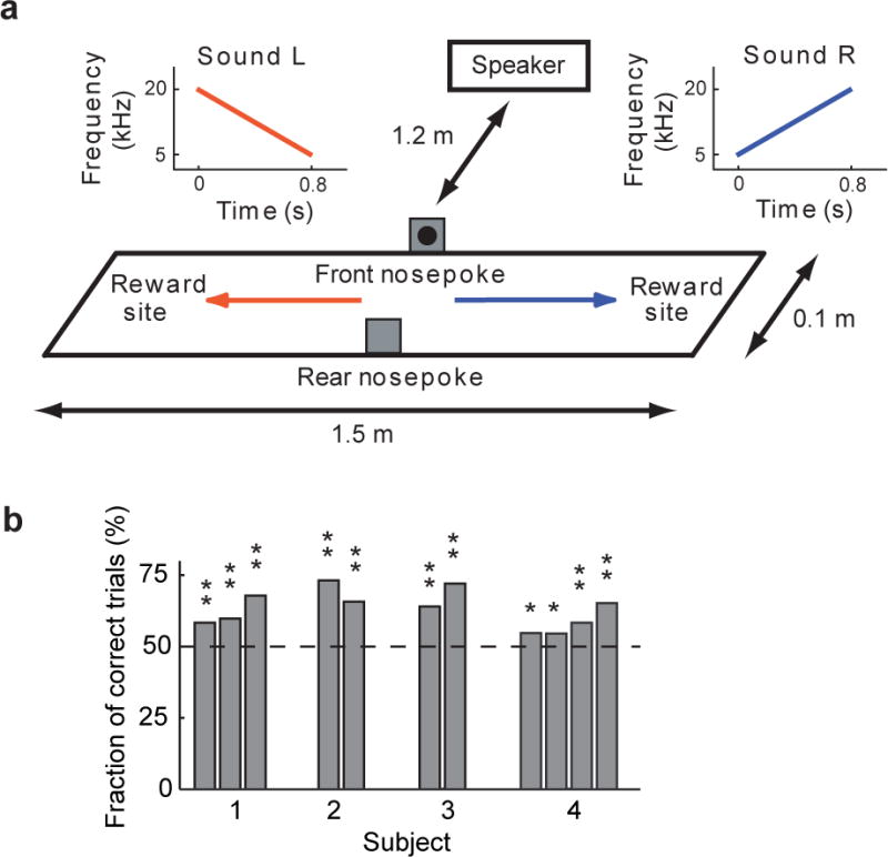
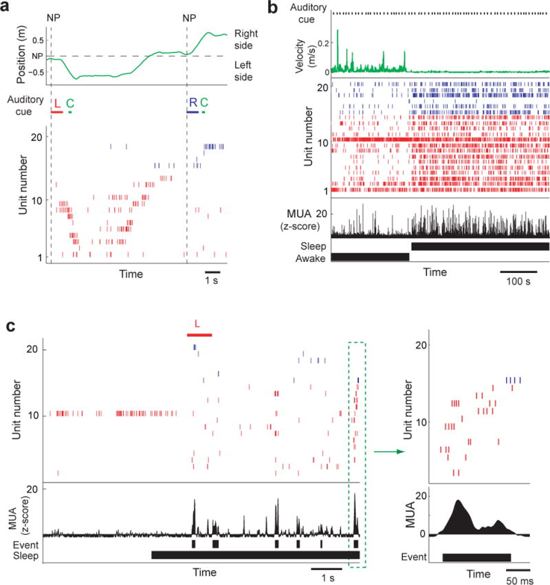
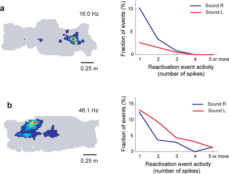
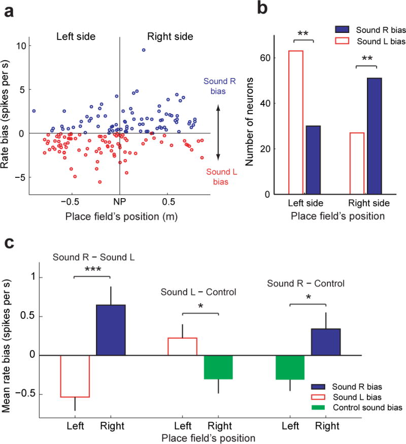
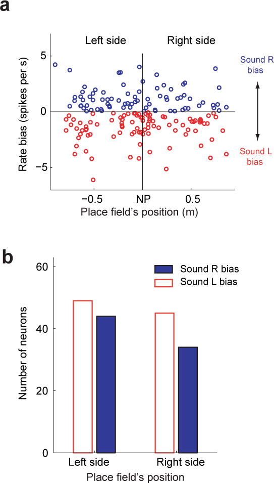
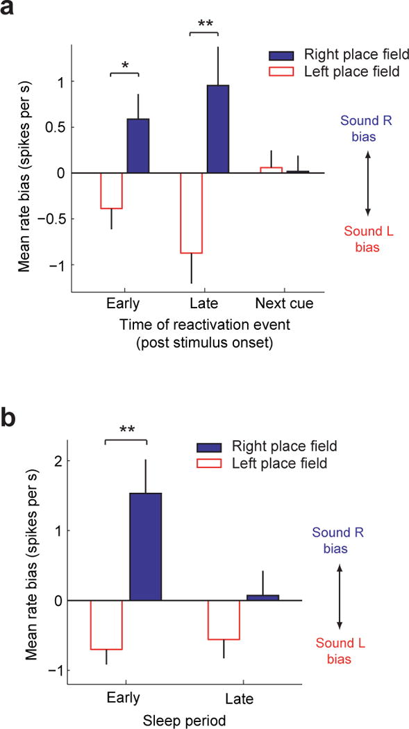
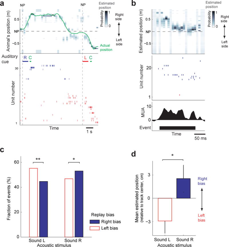
The hippocampus is essential for encoding self-experienced events into memory. During sleep, neural activity in the hippocampus related to a recent experience has been observed to spontaneously reoccur, and this 'replay' has been postulated to be important for memory consolidation. Task-related cues can enhance memory consolidation when presented during a post-training sleep session, and, if memories are consolidated by hippocampal replay, a specific enhancement for this replay should be observed. To test this, we trained rats on an auditory-spatial association task while recording from neuronal ensembles in the hippocampus. We found that, during sleep, a task-related auditory cue biased reactivation events toward replaying the spatial memory associated with that cue. These results indicate that sleep replay can be manipulated by external stimulation and provide further evidence for the role of hippocampal replay in memory consolidation.


Conflict of interest statement

The authors do not have any competing interests

Figures

Figure 1
Figure 1
Behavioral taska a. Behavioral task design. After the rat made a nosepoke, one of two sounds (Sound R or Sound L) was played from a speaker 1.2 m away from the track center (in front of the nosepoke). For a correct trial, the rat has to go the right side of the track for Sound R, and the left side of the track for Sound L. b. Performance on behavioral task. For the 11 recorded sessions in four subjects, task performance was above chance (* P < 0.05, ** P < 0.001, Binomial distribution).
Figure 2
Figure 2
Hippocampal activity during behavior and sleep (Rat 1, session 2) a. Neuronal ensemble activity during two correct behavioral trials- From top to bottom: (1) the spatial position (green) of the subject on the track, (2) acoustic stimuli- Sound R (R) in blue, a tone indicating a correct trial and reward delivery cue (C) in green, and Sound L (L) in red), (3) raster plot of place cell activity. Nosepokes (NP) are indicated by a vertical dashed line. Sound L was played when a nosepoke was made, and the rat correctly ran to the left side of the track. Having returned and made another nosepoke, the rat heard Sound R and ran to the right side of the track. In the raster plot, spikes from place cells with place fields on the right side of the track are blue, and left-sided place fields in red. Place fields are ordered from top to bottom by their location on the track (right → left side). b. Neuronal ensemble activity during the onset of sleep while the animal is resting in the sleep box after the behavioral task. From top to bottom (1) the onsets of acoustic stimuli, (2) animal’s velocity (green), (3) a raster plot of place cell activity (same format as in Fig. 2a), (4) multiunit activity, (5) awake/sleep classification. Note the increase in the time scale from Fig. 2a to Fig. 2b. c. Example of reactivation events occurring after the onset of sleep (from Fig. 2b). Prior to sleep onset, the rat was resting in the sleep chamber. The reactivation event in the green dashed box is shown to the right (same format as Fig. 2a). From top to bottom: (1) acoustic stimulus, (2) place cell activity (red=left-sided place fields, blue=right side place fields), (3) multiunit activity (MUA), (4) candidate reactivation events, (5) sleep classification.
Figure 3
Figure 3
Place cell responses during sleep reactivation events Place field (left) and plot of place cell activity (right) during reactivation events occurring after sound R (blue) and sound L (red). a. Rat 4, session 1, cluster 17 b. Rat 1, session 2, cluster 41
Figure 4
Figure 4
Rate bias during sleep reactivation events Error bars indicate the standard error of the mean (SEM). a. Rate bias during sleep replay events of individual place cells with place fields on the left or right side of the nosepoke (n=171). Place cells are ordered along the x-axis according to the location of their place field’s on the track. A positive rate bias indicates that the mean firing rate during replay events occurring after Sound R was higher than after Sound L. The vertical dashed line indicates the center of the track (location of the nosepoke). b. The number of place cells during sleep replay events with a Sound L or Sound R bias. Place cells are grouped according to the position of their place field along the track (left or right). Left-sided place cells had significantly more units with a Sound L bias than a Sound R bias (** P < 1.9 × 10⁻⁴, Binomial distribution). Opposite to this, right-sided place cells had significantly more units with a Sound R bias than a Sound L bias (** P < 0.003, Binomial distribution). c. The mean rate bias for left-sided and right-sided place cells during sleep replay events. In the first column, the mean rate bias compares Sound R and Sound L. In the second column the mean rate bias compares Sound L and the two task-related control sounds (reward and error cue). In the third column the mean rate bias compares Sound R and the two task-related control sounds (reward and error cue). The mean rate bias is significantly different between place fields on the left side and right side of the track in all three analyses (1-way ANOVA: Sound R − Sound L, *** P < 7.8 × 10⁻⁵; Sound L − control, * P < 0.04; Sound R − control, * P < 0.02).
Figure 5
Figure 5
Rate bias during awake reactivation events Error bars indicate the standard error of the mean (SEM). a. Rate bias of individual place cells during awake reactivation events (n = 172). Format of plot is the same as Fig 4a. Pearson correlation coefficient: r = –0.03, P = 0.74 b. Number of neurons with sound R and sound L biases (Binomial test, left-sided place fields: P=0.27; right-sided place fields: P=0.87)
Figure 6
Figure 6
Temporal dynamics of sound-evoked reactivation bias Error bars indicate the standard error of the mean (SEM). a. Mean rate bias for events occurring at different time points relative to the onset of the acoustic stimulus. Events were divided into three groups based on when the events occurred, relative to the sound. For the first group (Early), events occurred between 0–5.4 seconds after the onset of the sound (1-way ANOVA, * P < 0.007). For the second group (Late), events occurred between 5.4–10.8 seconds after the onset of the sound, but before the onset of the next acoustic stimulus (1-way ANOVA, ** P < 7.4 × 10⁻⁴). For the third group, events occurred after the onset of the next acoustic stimulus (1-way ANOVA, P = 0.87). b. In each experiment, the sleep session consisting of one or more sleep/wake cycles was concatenated, and divided into two periods, first half (Early) and second half (Late) of the total time spent asleep. The mean rate bias was calculated using all reactivation events in the Early and Late sleep periods. A significant mean rate bias difference between right and left sided place fields was observed during the Early period of sleep (1-way ANOVA, **P< 1.7 × 10⁻⁵).
Figure 7
Figure 7
Bias in Bayesian decoded position during sleep reactivation events a. Example of Bayesian decoding during two behavioral trials- From top to bottom: (1) the estimated spatial position (Bayesian reconstruction) and actual position (green) of the subject on the track, (2) acoustic stimuli- Sound R (R) in blue, a tone indicating a correct trial and reward delivery cue (C) in green, and Sound L (L) in red), (3) raster plot of place cell activity. Nosepokes (NP) are indicated by a vertical dashed line. All place fields with peak firing rates greater then 0.5 spk/s are displayed in the raster plot, and subsequently used for the reconstruction. Sound R was played when a nosepoke was made, and the rat correctly ran to the right side of the track. Having returned and made another nosepoke, the rat heard Sound L and ran to the left side of the track. In the raster plot, spikes from place cells with place fields on the right side of the track are blue, and left-sided place fields in red. Place fields are ordered from top to bottom by their location on the track (right → left side). b. Example of Bayesian decoding of a reactivation event during sleep (Rat 2, session 1). From top to bottom: (1) the estimated spatial position of the replay (Bayesian reconstruction), (2) place cell activity (red=left-sided place fields, blue=right side place fields), (3) multiunit activity (MUA), (4) reactivation event detection. c. Fraction of replay events occurring after Sound R and Sound L with a mean decoded position with a right or left-sided bias. Sound L had significantly more replay events with a left-sided bias (** P < 0.002, Binomial distribution), while Sound R was significantly biased towards the right side (* P < 0.05, Binomial distribution). d. Mean estimated position during replay events occurring after Sound L and Sound R. Sound R replay events have a positive bias (mean position is on the right side of the track) while Sound L replay events have a negative bias (mean position is on the left side of the track). The difference between the mean estimate position for Sound L and Sound R replay events was statistically significant (* P < 0.006, Wilcoxon rank sum test). The mean estimated track position is relative to the center of the track (negative values- left side, positive values- right side). From the track center (location of nosepoke), the track extends 75 cm to each side. Error bars indicate the standard error of the mean (SEM).

Comment in

  • Sleep tight, wake up bright.
    Kelemen E, Born J. Kelemen E, et al. Nat Neurosci. 2012 Oct;15(10):1327-9. doi: 10.1038/nn.3227. Nat Neurosci. 2012. PMID: 23007185 No abstract available.

References

References for Online Methods

    1. Davidson TJ, Kloosterman F, Wilson MA. Hippocampal replay of extended experience. Neuron. 2009;63(4):497–507. - PMC - PubMed
    1. Hirase H, Leinekugel X, Czurkó A, Csicsvari J, Buzsáki G. Firing rates of hippocampal neurons are preserved during subsequent sleep episodes and modified by novel awake experience. Proc Natl Acad Sci. 2001;98(16):9386–90. - PMC - PubMed
    1. Ji D, Wilson MA. Coordinated memory replay in the visual cortex and hippocampus during sleep. Nat Neurosci. 2007;10(1):100–7. - PubMed
    1. Lansink CS, Goltstein PM, Lankelma JV, Joosten RN, McNaughton BL, Pennartz CM. Preferential reactivation of motivationally relevant information in the ventral striatum. J Neurosci. 2008;28(25):6372–82. - PMC - PubMed
    1. Zhang K, Ginzburg I, McNaughton BL, Sejnowski TJ. Interpreting neuronal population activity by reconstruction: unified framework with application to hippocampal place cells. J Neurophysiol. 1998;79(2):1017–44. - PubMed

References

    1. Dupret D, O’Neill J, Pleydell-Bouverie B, Csicsvari J. The reorganization and reactivation of hippocampal maps predict spatial memory performance. Nat Neurosci. 2010;13(8):995–1002. - PMC - PubMed
    1. Squire LR. Memory and the hippocampus: a synthesis from findings with rats, monkeys, and humans. Psychol Rev. 1992;99(2):195–231. - PubMed
    1. Marshall L, Born J. The contribution of sleep to hippocampus-dependent memory consolidation. Trends Cogn Sci. 2007;11(10):442–50. - PubMed
    1. Siapas AG, Wilson MA. Coordinated interactions between hippocampal ripples and cortical spindles during slow-wave sleep. Neuron. 1998;21(5):1123–8. - PubMed
    1. Sirota A, Csicsvari J, Buhl D, Buzsáki G. Communication between neocortex and hippocampus during sleep in rodents. Proc Natl Acad Sci U S A. 2003;100(4):2065–9. - PMC - PubMed

Publication types