

doi: 10.1038/nmeth.2177.
Epub 2012 Sep 2.
Coupling endonucleases with DNA end-processing enzymes to drive gene disruption
Affiliations
- PMID: 22941364
- PMCID: PMC3602999
- DOI: 10.1038/nmeth.2177
Item in Clipboard
Coupling endonucleases with DNA end-processing enzymes to drive gene disruption
Nat Methods.
2012 Oct.
Abstract
Targeted DNA double-strand breaks introduced by rare-cleaving designer endonucleases can be harnessed for gene disruption applications by engaging mutagenic nonhomologous end-joining DNA repair pathways. However, endonuclease-mediated DNA breaks are often subject to precise repair, which limits the efficiency of targeted genome editing. To address this issue, we coupled designer endonucleases to DNA end-processing enzymes to drive mutagenic break resolution, achieving up to 25-fold enhancements in gene disruption rates.
Figures
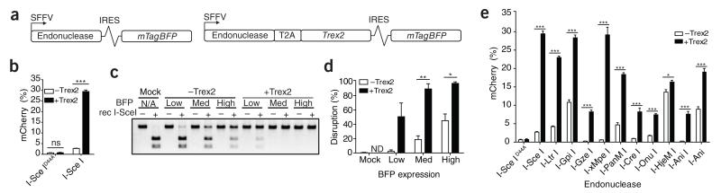
Coupling endonucleases to exonucleases increases gene disruption. (a) Schematic of Trex2 exonuclease expression vectors. SFFV, spleen focus-forming virus promoter/enhancer; IRES, internal ribosomal entry site. (b) Quantification of mCherry expression in BFP-positive HEK293T cells transfected with the indicated vectors and analyzed 72 h after transfection (n = 3). (c) Sce-digestion NHEJ assay to measure total gene disruption. Cells were sorted based on the gates indicated in supplementary figure 2b. Rec, recombinant. (d) Quantification of band intensity (undigested out of total) of three independent experiments of the Sce digestion assay as performed in c. ND, not determined. (e) Quantification of mCherry expression in BFP-positive HEK293T cells harboring the respective TLR target for each of the indicated homing endonucleases (n = 3) and analyzed 72 h after transfection with the indicated vectors. Error bars, s.e.m. P values (*P < 0.05, **P < 0.005 and ***P < 0.0005) were calculated using the Student’s two-tailed unpaired t-test to compare the samples indicated in this and all subsequent figures.
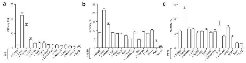
DNA end-processing enzymes library screen. (a–c) Quantification of mCherry expression in BFP-positive HEK293T cells harboring the Sce-TLR (a; n = 5 or 6), the TLR with the CCR5 TALEN target site (b, n = 3) or the TLR with the VF2468 ZFN target site (c, n = 3). All quantifications were done at 72 h after transfection with the indicated vectors. Endonuclease expression was in all cases tracked with BFP. Asterisks indicate a significant difference between samples expressing an enzyme from the library with designer endonuclease and endonuclease with empty vector alone. Error bars, s.e.m.
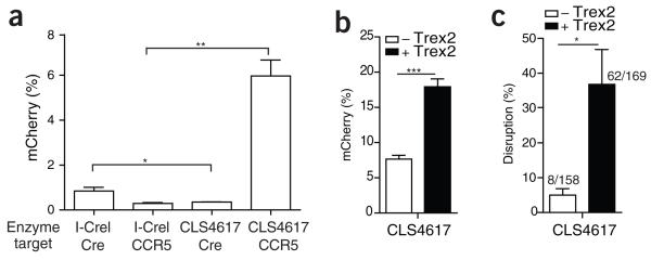
Trex2 increases knockout of endogenous CCR5 with an engineered homing endonuclease. (a) Quantification of mCherry expression in BFP-positive HEK293T cells containing the TLR with the I-CreI cognate and CLS4617 targets 72 h after transfection with the indicated enzyme (n = 3). (b) Quantification of mCherry expression in BFP-positive HEK293T TLR cells with the CLS4617 target 72 h after transfection with the indicated vectors (n = 3). (c) Quantification of total disruption rate, analyzed by sequencing the CCR5 TLR target of BFP-sorted CD34+ cells transduced with the indicated lentiviral vectors at a multiplicity of infection of 20 (n = 3). Numbers above bars indicate total number of sequences (mutated/total). Error bars, s.e.m..
References
Publication types
MeSH terms
Substances
Grants and funding
- T32 GM07270/GM/NIGMS NIH HHS/United States
- U19 AI096111/AI/NIAID NIH HHS/United States
- U19-AI96111/AI/NIAID NIH HHS/United States
- RL1 CA133832/CA/NCI NIH HHS/United States
- T32 GM007270/GM/NIGMS NIH HHS/United States
- PL1-HL092557/HL/NHLBI NIH HHS/United States
- PL1 HL092557/HL/NHLBI NIH HHS/United States
- UL1 DE019582/DE/NIDCR NIH HHS/United States
- RL1CA133832/CA/NCI NIH HHS/United States
- R01-HL075453/HL/NHLBI NIH HHS/United States
- RL1-HL92554/HL/NHLBI NIH HHS/United States
- R01 HL075453/HL/NHLBI NIH HHS/United States
- UL1 RR024921/RR/NCRR NIH HHS/United States
- RL1-HL092553/HL/NHLBI NIH HHS/United States
- UL1DE019582/DE/NIDCR NIH HHS/United States
- RL1 HL092553/HL/NHLBI NIH HHS/United States
- R41 GM085876/GM/NIGMS NIH HHS/United States
- RL1 HL092554/HL/NHLBI NIH HHS/United States
LinkOut - more resources
Full Text Sources
Other Literature Sources
Research Materials
