

Social inclusion enhances biological motion processing: a functional near-infrared spectroscopy study
- PMID: 22941501
- PMCID: PMC3537903
- DOI: 10.1007/s10548-012-0253-y
Social inclusion enhances biological motion processing: a functional near-infrared spectroscopy study
Erratum in
-
Erratum to: Social Inclusion Enhances Biological Motion Processing: A Functional Near-Infrared Spectroscopy Study.Brain Topogr. 2015 Jan;28(1):184-5. doi: 10.1007/s10548-014-0419-x. Brain Topogr. 2015. PMID: 25466467 No abstract available.
Abstract
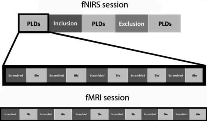
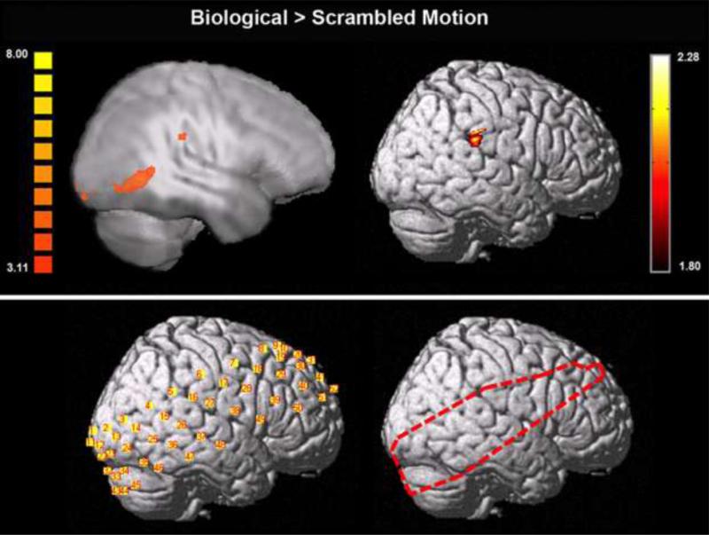
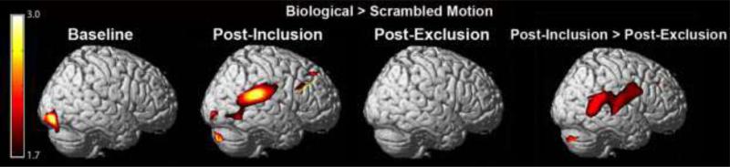
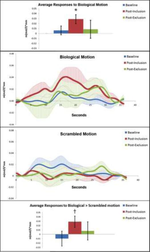
Humans are especially tuned to the movements of other people. Neural correlates of this social attunement have been proposed to lie in and around the right posterior superior temporal sulcus (STS) region, which robustly responds to biological motion in contrast to a variety of non-biological motions. This response persists even when no form information is provided, as in point-light displays (PLDs). The aim of the current study was to assess the ability of functional near-infrared spectroscopy (fNIRS) to reliably measure brain responses to PLDs of biological motion, and determine the sensitivity of these responses to interpersonal contextual factors. To establish reliability, we measured brain activation to biological motion with fNIRS and functional magnetic resonance imaging (fMRI) during two separate sessions in an identical group of 12 participants. To establish sensitivity, brain responses to biological motion measured with fNIRS were subjected to an additional social manipulation where participants were either socially included or excluded before viewing PLDs of biological motion. Results revealed comparable brain responses to biological motion using fMRI and fNIRS in the right supramarginal gyrus. Further, social inclusion increased brain responses to biological motion in right supramarginal gyrus and posterior STS. Thus, fNIRS can reliably measure brain responses to biological motion and can detect social experience-dependent modulations of these brain responses.
Figures
References
-
- Allison T, Puce A, McCarthy G. Social Perception from visual cues: Role of the STS region. Trends Cogn Sci. 2000;4:267–278. doi:10.1016/S1364-6613(00)01501-1. - PubMed
-
- Baumeister RF, Leary MR. The need to belong: Desire for interpersonal attachments as a fundamental human motivation. Psychol Bull. 1995;117:497–529. doi: 10.1037/0033-2909.117.3.497. - PubMed
-
- Bidet-Ildei C, Chauvin A, Coello Y. Observing or producing a motor action improves later perception of biological motion: Evidence for a gender effect. Acta Psychol. 2010;134:215–224. doi:10.1016/j.actpsy.2010.02.002. - PubMed
-
- Blake R, Shiffrar M. Perception of human motion. Annu Rev Psychol. 2007;58:47–73. doi:10.1146/annurev.psych.57.102904.190152. - PubMed
Publication types
MeSH terms
Grants and funding
LinkOut - more resources
Full Text Sources
Medical
