. 2012 Nov 1;40(20):10312-23.
doi: 10.1093/nar/gks807. Epub 2012 Aug 31.

PALB2 self-interaction controls homologous recombination

Affiliations

PALB2 self-interaction controls homologous recombination

Rémi Buisson et al. Nucleic Acids Res. .

Abstract

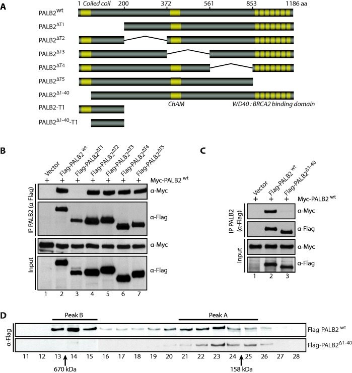
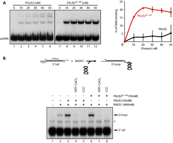
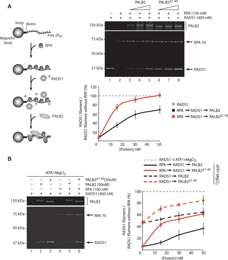
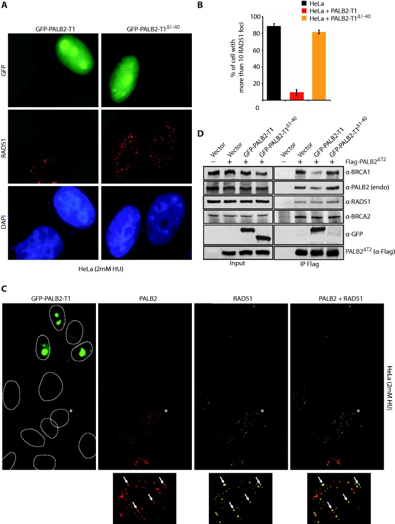
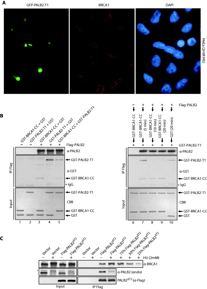
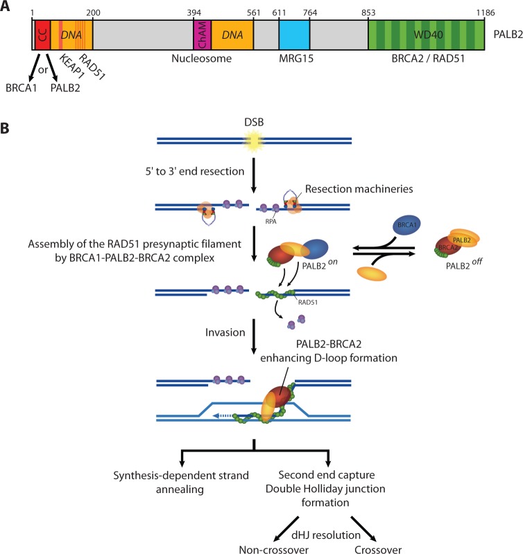
PALB2 is essential for BRCA2 anchorage to nuclear structures and for homologous recombinational repair of DNA double-strand breaks. Here, we report that the N-terminal coiled-coil motif of PALB2 regulates its self-association and homologous recombination. Monomeric PALB2 shows higher efficiency to bind DNA and promotes RAD51 filament formation with or without the inhibitory effect of Replication Protein A. Moreover, overexpression of the PALB2 coiled-coil domain severely affects RAD51 loading to DNA damage sites suggesting a competition between PALB2 self-interaction and PALB2-BRCA1 interaction. In the presence of DNA damage, the switch between PALB2-PALB2 and PALB2-BRCA1 interactions allows the activation of HR. Controlling HR via PALB2 self-interactions could be important to prevent aberrant recombination in normal conditions and activate DNA repair when required.


Figures

Figure 1.
Figure 1.
PALB2 contains a self-interaction domain localized at its N-terminus. (A) Schematic representation of PALB2 and PALB2 truncations used in this study. (B–C) Myc-PALB2 and Flag-PALB2 (PALB2 wild-type or PALB2 deletion mutants) were co-expressed in HEK293T cells and followed by an IP with anti-Flag antibody. Immunoprecipitated proteins were detected by western blotting with anti-Flag or anti-Myc antibodies. (D) Size-exclusion chromatography analysis of HEK293T whole-cell extracts expressing Flag-PALB2 or Flag-PALB2Δ1–40. Proteins of each fraction were detected by western blotting with an anti-Flag antibody. Molecular weight standards are indicated at the bottom of the Figure.
Figure 2.
Figure 2.
DNA binding by monomeric PALB2 and D-loop formation. (A) PALB2 or PALB2Δ1–40 bind ssDNA. Electrophoretic mobility shift assays were performed with PALB2 or PALB2Δ1–40 (10–50 nM) and 60 nt ssDNA (100 nM nucleotides) and analyzed on a 8% polyacrylamidegel. Right: quantification of the results. (B) Top: schematic of D-loop assay. D-loop reactions were performed with RAD51 alone (lane 2) or combinations of RAD51 and PALB2 or PALB2Δ1–40 (lanes 3–8). The band indicated with an asterisk corresponds to annealed tailed molecules. CCC designates Covalently Closed Circular supercoiled plasmid.
Figure 3.
Figure 3.
Stimulation of RAD51 filament formation by monomeric PALB2. (A) Left: schematic of RPA displacement assay. Right: RPA bound to a ssDNA oligonucleotide prevents RAD51 assembly (lane 2) whereas addition of PALB2 or PALB2Δ1–40 (15, 30 and 50 nM) stimulates RAD51 filament formation in presence of RPA (lanes 3–8). Bottom: quantification of the results. The Y-axis represents the ratio between the values obtained for RAD51 filament in the presence of RPA and PALB2 or PALB2Δ1–40 divided by the value obtained for RAD51 filament with ATP and magnesium without RPA (set at 100%). (B) Left: RPA displacement assay in the absence of ATP and MgCl2. Right: quantification of the results. The Y-axis represents the ratio between the values obtained for RAD51 filament in the absence of ATP/MgCl2 with or without RPA and PALB2 or PALB2Δ1–40 divided by the value obtained for RAD51 filament with ATP and magnesium without RPA (set at 100%).
Figure 4.
Figure 4.
Inhibition of RAD51 foci formation by the PALB2 coiled-coil domain. (A) Expression in Hela cells of GFP-PALB2-T1 with or without the coiled-coil domain (GFP-PALB2-T1Δ1–40). Immunofluorescence pictures with anti-GFP and anti-RAD51 are shown. (B) Quantification of the experiment shown in (A). (C) Expression in Hela cells of GFP-PALB2-T1 followed by immunofluorescence with anti-PALB2 (in red) and anti-RAD51 (in Far-red). The dotted lines delimit the DAPI staining. The cell indicated with an asterisk was enlarged at the bottom of the figure and arrows indicate a co-localization between PALB2 and RAD51 foci. (D) HEK293T cells were transfected with a vector alone or Flag-PALB2ΔT2, with GFP-PALB2-T1 or GFP-PALB2-T1Δ1–40. Flag-IPs were conducted, followed by western blotting with the indicated antibodies.
Figure 5.
Figure 5.
(A) Expression of GFP-PALB2-T1 does not affect BRCA1 foci formation. Immunofluorescence staining was performed with anti-BRCA1 (red). The DAPI staining is also shown. (B) Purified GST-BRCA1-CC competes with PALB2 self-interaction. Proteins were incubated with PALB2-Flag/His followed by IP with anti-Flag and western blotting with anti-GST and anti-PALB2 antibodies. Left: the order of addition of each protein is shown on the top of the gel. The proteins stained by Coomassie brilliant blue (CBB) are also shown. (C) HEK293T cells were transfected with an empty vector or a vector containing Flag-PALB2ΔT2, treated with HU (2 mM, 20 h) and Flag-PALB2ΔT2 was immunoprecipitated with an anti-Flag antibody. Immunoprecipitated proteins were detected by western blotting with the indicated antibodies. Quantification of the increase is estimated by a titration of Flag-PALB2ΔT2 + HU IP. The band indicated with an asterisk corresponds to the phosphorylated form of PALB2.
Figure 6.
Figure 6.
(A) Schematic representation of PALB2 domains. (B) Model for the role of PALB2 during HR repair (see main text for description).

References

    1. Ciccia A, Elledge SJ. The DNA damage response: making it safe to play with knives. Mol. Cell. 2010;40:179–204. - PMC - PubMed
    1. Moynahan ME, Jasin M. Mitotic homologous recombination maintains genomic stability and suppresses tumorigenesis. Nat. Rev. Mol. Cell Biol. 2010;11:196–207. - PMC - PubMed
    1. San Filippo J, Sung P, Klein H. Mechanism of eukaryotic homologous recombination. Ann. Rev. Biochem. 2008;77:229–257. - PubMed
    1. Davies AA, Masson JY, McIlwraith MJ, Stasiak AZ, Stasiak A, Venkitaraman AR, West SC. Role of BRCA2 in control of the RAD51 recombination and DNA repair protein. Mol. Cell. 2001;7:273–282. - PubMed
    1. Carreira A, Hilario J, Amitani I, Baskin RJ, Shivji MK, Venkitaraman AR, Kowalczykowski SC. The BRC repeats of BRCA2 modulate the DNA-binding selectivity of RAD51. Cell. 2009;136:1032–1043. - PMC - PubMed

Publication types

MeSH terms