. 2013 Feb;57(2):763-74.
doi: 10.1002/hep.26042. Epub 2013 Jan 10.

Beta-catenin-NF-κB interactions in murine hepatocytes: a complex to die for

Affiliations

Beta-catenin-NF-κB interactions in murine hepatocytes: a complex to die for

Kari Nejak-Bowen et al. Hepatology. 2013 Feb.

Abstract

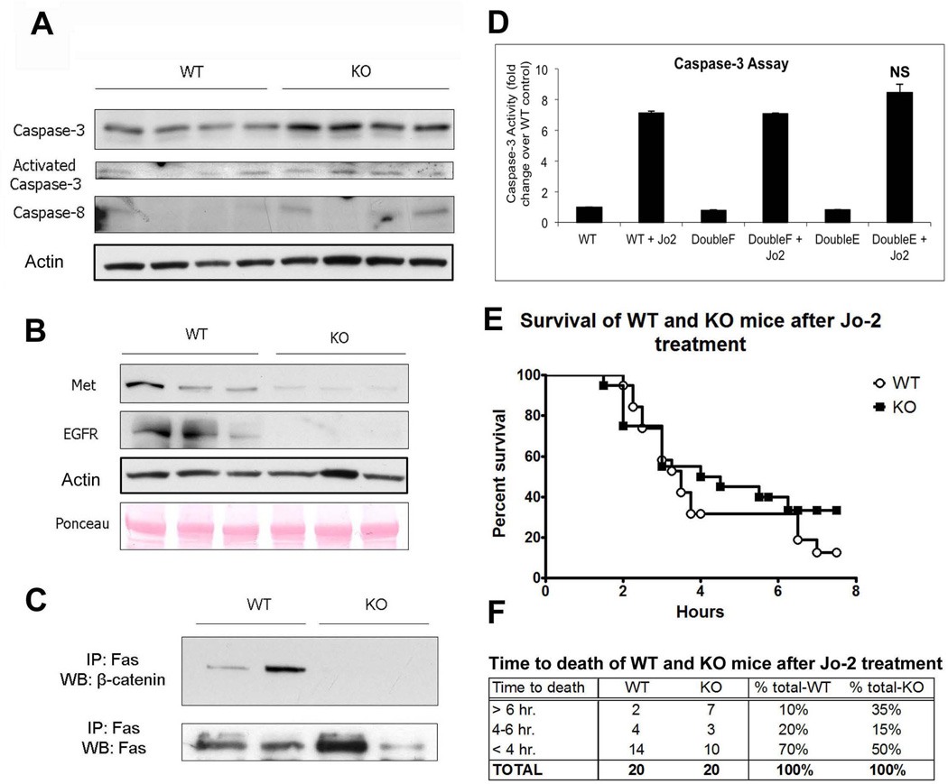
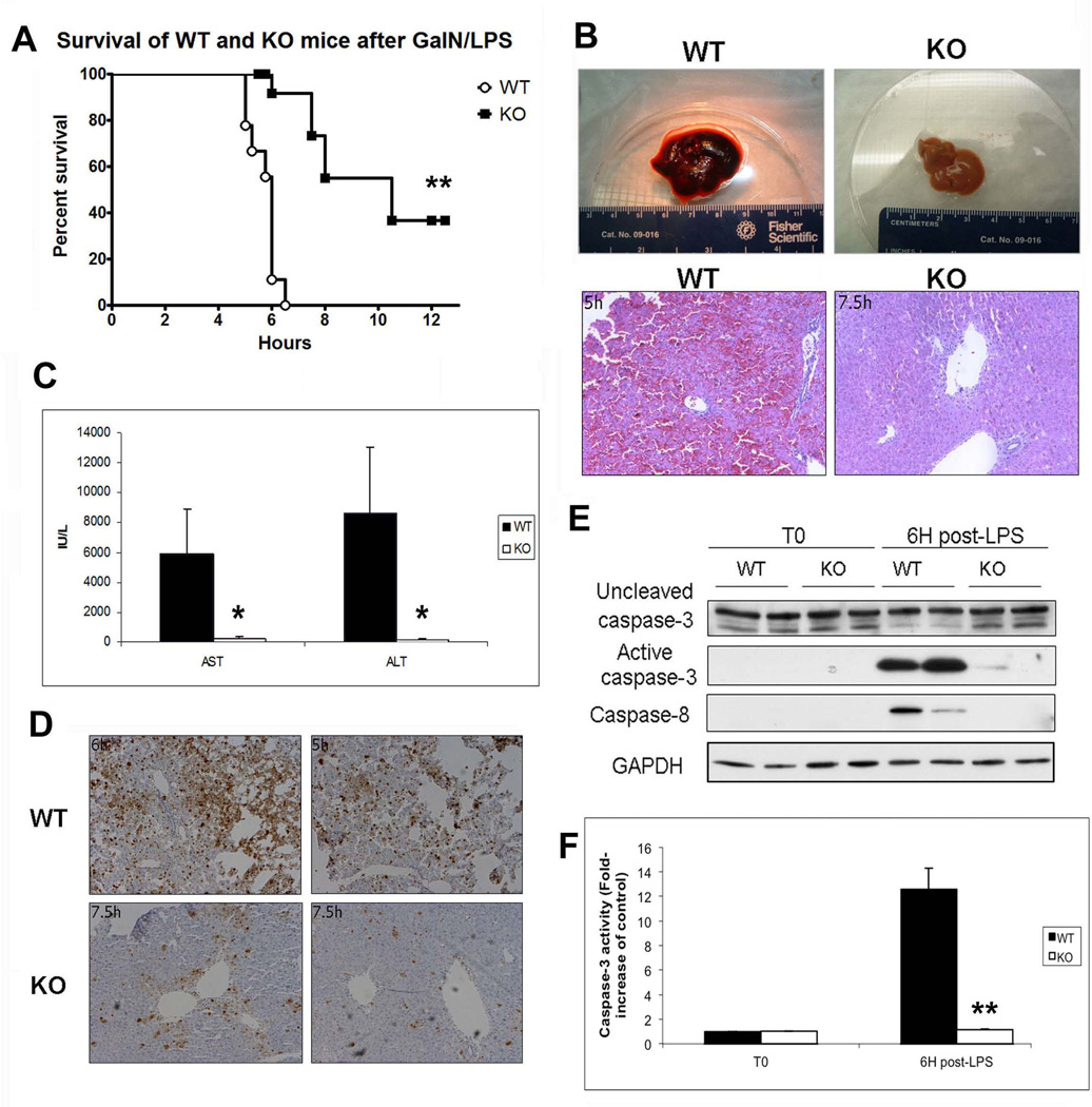
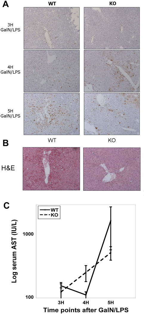
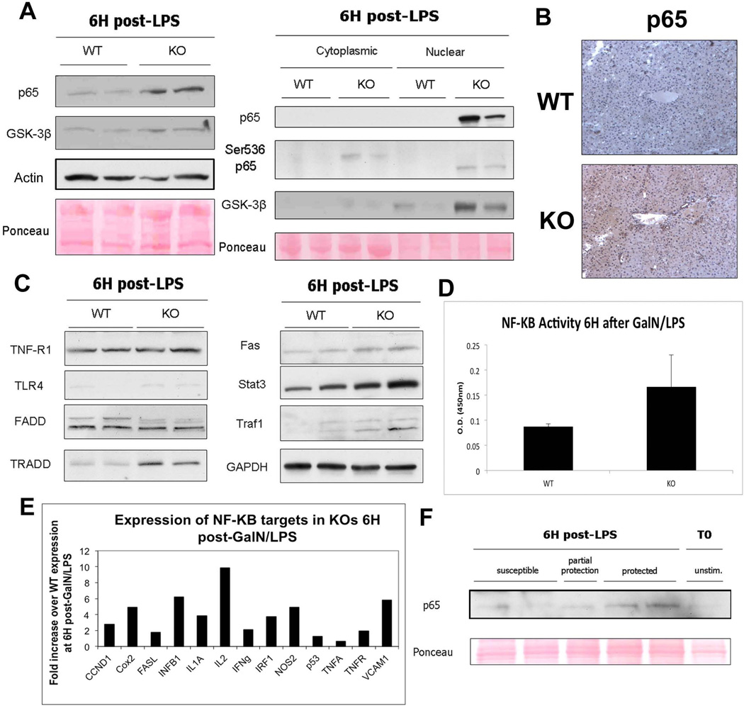
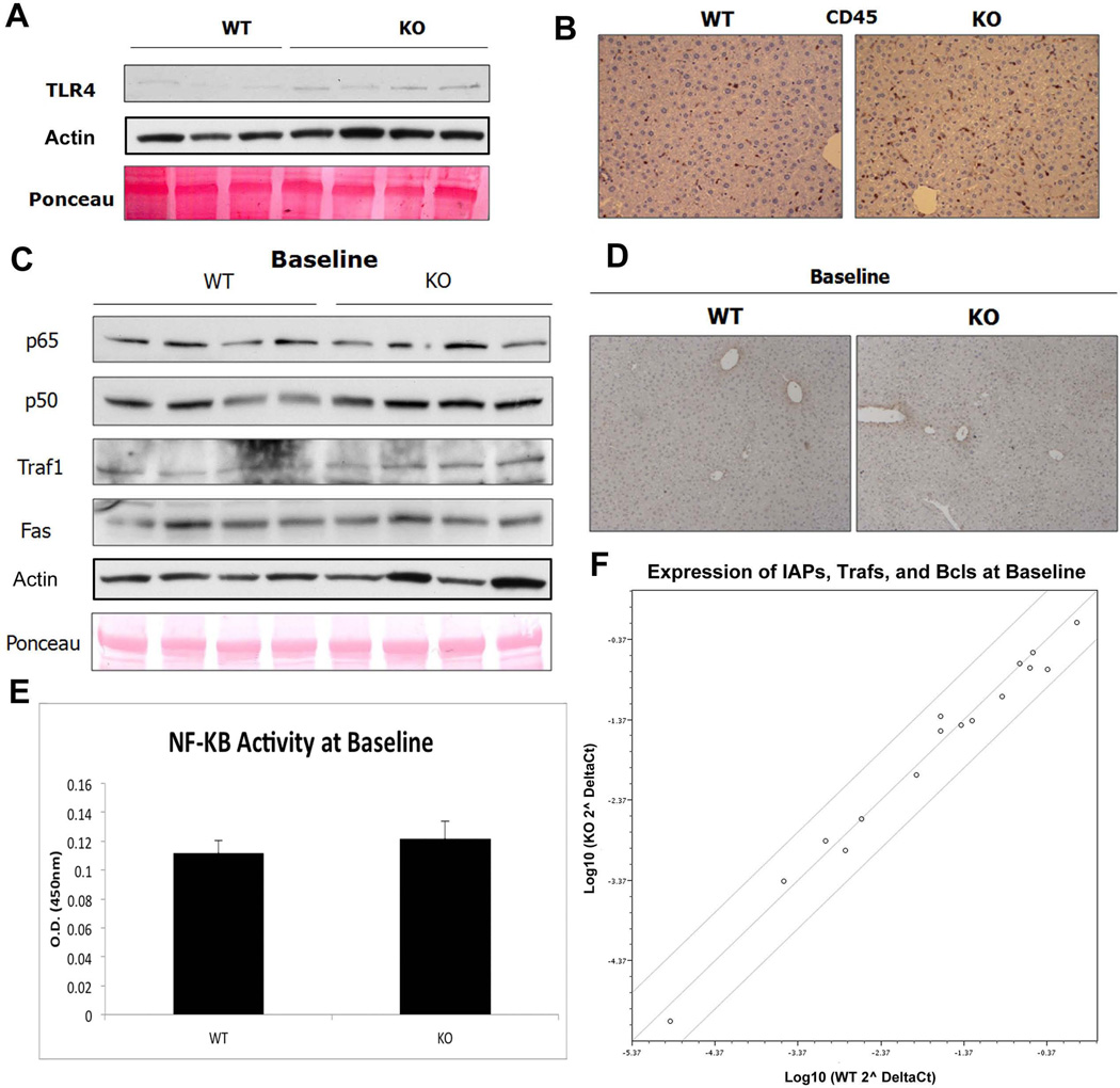
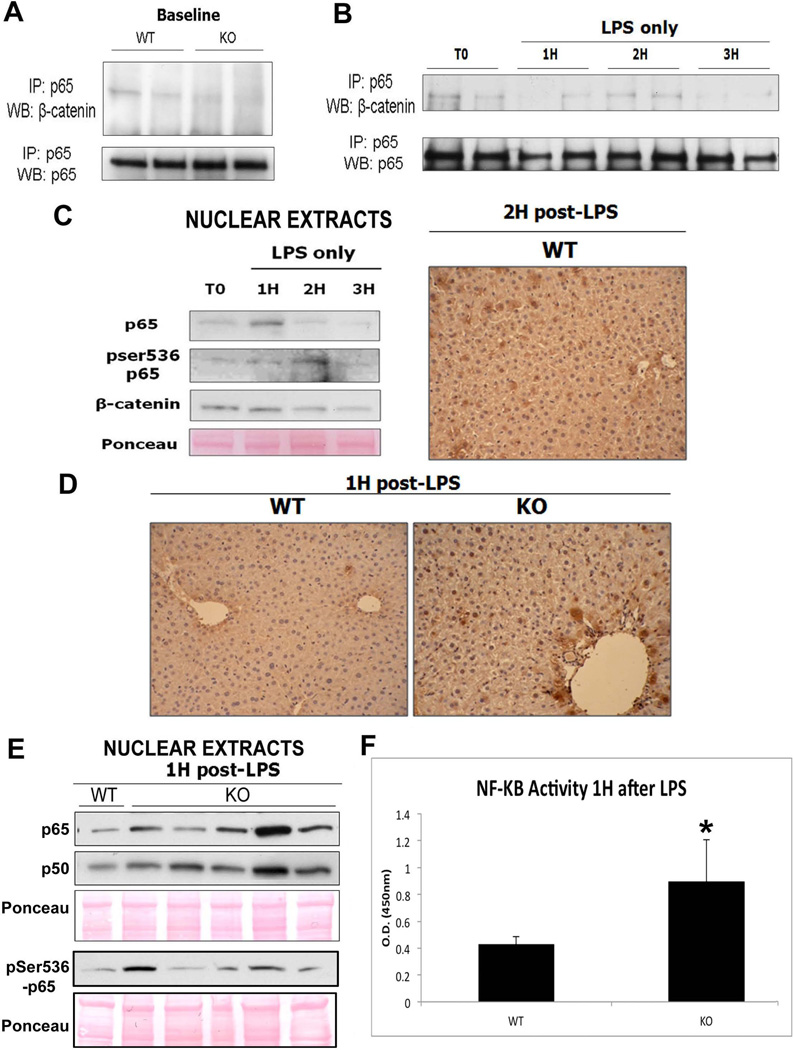
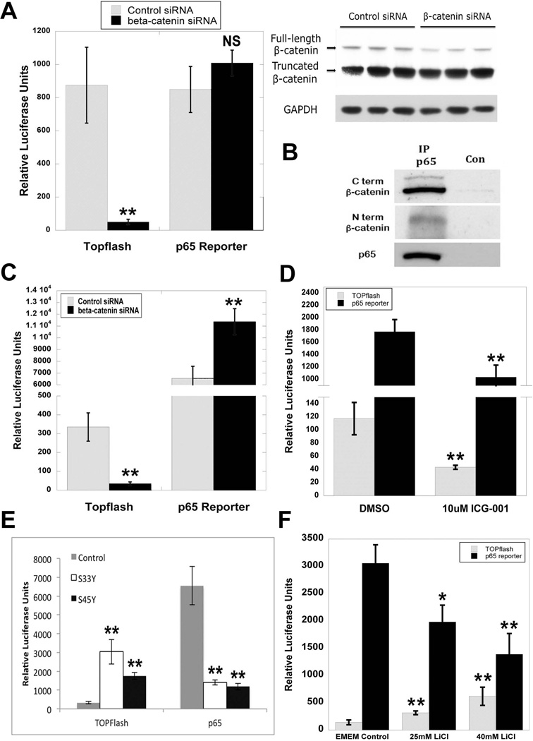
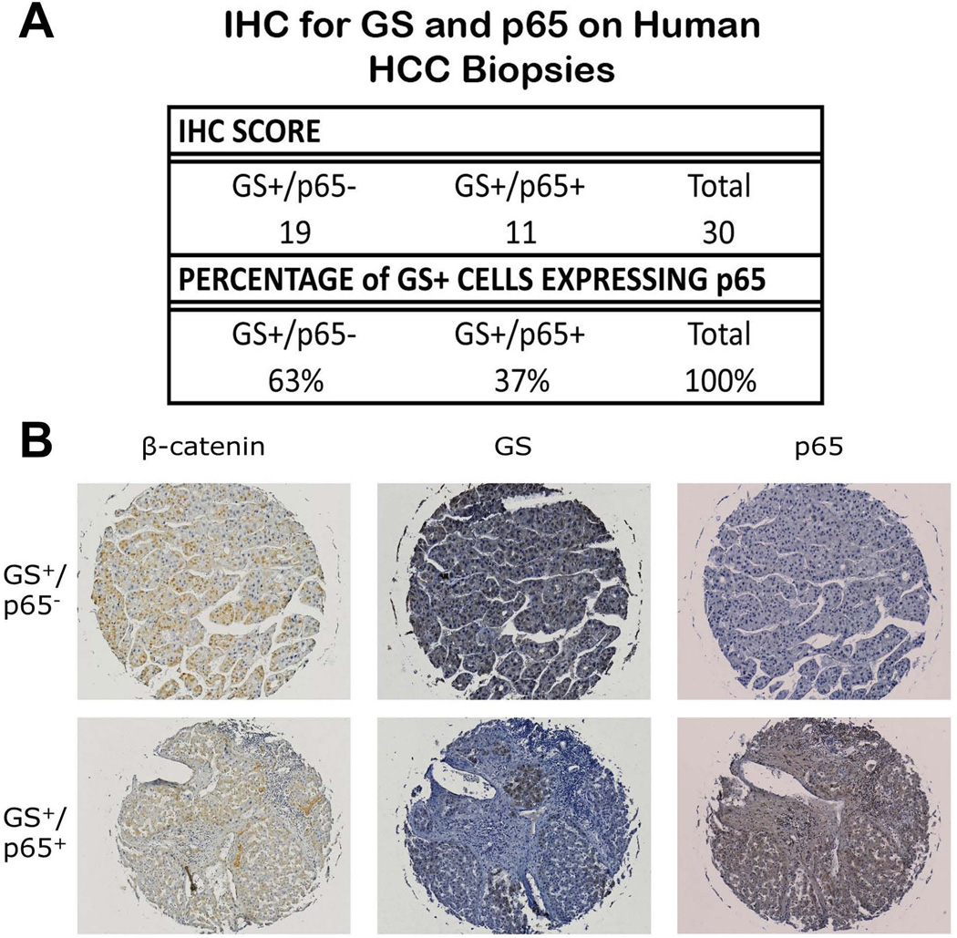
Wnt/β-catenin signaling plays an important role in hepatic homeostasis, especially in liver development, regeneration, and cancer, and loss of β-catenin signaling is often associated with increased apoptosis. To elucidate how β-catenin may be regulating hepatocyte survival, we investigated the susceptibility of β-catenin conditional knockout (KO) mice and their wild-type (WT) littermates to Fas and tumor necrosis factor-α (TNF-α), two common pathways of hepatocyte apoptosis. While comparable detrimental effects from Fas activation were observed in WT and KO, a paradoxical survival benefit was observed in KO mice challenged with D-galactosamine/lipopolysaccharide. KO mice showed significantly lower morbidity and liver injury due to early, robust, and protracted activation of NF-κB in the absence of β-catenin. Enhanced NF-κB activation in KO mice was associated with increased basal inflammation and Toll-like receptor 4 expression and lack of the p65/β-catenin complex in hepatocytes. The p65/β-catenin complex in WT livers underwent temporal dissociation allowing for NF-κB activation to regulate hepatocyte survival following TNF-α-induced hepatic injury. Decrease of total β-catenin protein but not its inactivation induced p65 activity, whereas β-catenin stabilization either chemically or due to mutations repressed it in hepatomas in a dose-dependent manner, whereas β-catenin stabilization repressed it either chemically or due to mutations.

Conclusion: The p65/β-catenin complex in hepatocytes undergoes dynamic changes during TNF-α-induced hepatic injury and plays a critical role in NF-κB activation and cell survival. Modulation of β-catenin levels is a unique mode of regulating NF-κB activity and thus may present novel opportunities in devising therapeutics in specific hepatic injuries.


Conflict of interest statement

Disclosure: None of the authors have any potential conflicts of interest pertaining to the current manuscript.

Figures

Figure 1
Figure 1. β-catenin KO animals show alterations in components of the Fas pathway, which does not result in increased susceptibility to Fas-mediated apoptosis
(A) Caspase protein expression and activity is higher in KOs than in WTs at baseline; actin represents loading control. (B) Expression of Met and EGFR is decreased in KOs compared to WTs at baseline, as assessed by WB. (C) IP shows that Fas and β-catenin associate strongly in WT livers at baseline and that this association is absent in KO livers. (D) Active caspase-3 activity is not significantly changed in Hepa 1–6 cells transfected with either WT, constitutively active, or inactive β-catenin followed by Jo-2 treatment, as measured by fluorometric assay. (E) Kaplan-Meier survival analysis of WT and KO mice (n=20) after i.v. injection with Jo-2 shows no significant differences in overall survival between the two groups. (F) Grouping WT and KO animals treated with Jo-2 by time of death shows that KO animals are slightly more resistant to Fas-mediated apoptosis than WT animals.
Figure 2
Figure 2. β-catenin KO mice are protected from injury induced via the TNF-α pathway
(A) Kaplan-Meier survival analysis of WT and KO mice after treatment with GalN/LPS shows that KOs survive significantly longer than their WT counterparts (n≥9; **P<0.01). (B) Gross liver specimens from WT and KO mice injected with GalN/LPS demonstrate that WT livers become engorged with blood 6H post-injection, while the KO livers harvested at the same time appear normal. Corresponding histology is characteristic of massive inflammation and apoptotic cell death in WT livers and near normal histology in KO livers as shown by H&E. (C) Serum AST and ALT levels are significantly higher in WT mice than in KO mice 6H after GalN/LPS treatment (n=3; *P<0.05). (D) The number of apoptotic hepatocytes 6H after GalN/LPS injection (TUNEL IHC) is dramatically lower in KO mice as compared to WT controls. (E) Cleaved caspase-3 and caspase-8 is increased in WTs compared to KOs after 6H of GalN/LPS treatment, as shown in WB. GAPDH represents loading control. (F) The level of active caspase-3 activity is increased in WTs after 6H of GalN/LPS compared to KOs as measured by fluorometric assay (n=3; **P<0.01).
Figure 3
Figure 3. Progressive damage to WT livers after GalN/LPS, while damage in KO livers is arrested after an early onset
(A) KO livers have more TUNEL-positive cells compared to WT at 4H post-GalN/LPS. At 5H there is a rapid increase in the number of apoptotic cells in the WT compared to the KOs, which surpasses KO, while KO show no progression in the numbers of TUNEL-positive cells. (B) WTs have significantly more damage than KOs as early as 5H after GalN/LPS, as shown by H&E staining. (C) Serum AST levels are increased in KOs at 4H, while at 5H the AST in WTs has surpassed that seen in the KOs.
Figure 4
Figure 4. Cytoprotective proteins and expression of downstream NF-κB targets are higher in KO livers than in WT livers 6H post-GalN/LPS
(A) WB show that both p65 and GSK-3β are increased in whole-liver lysates from KOs as compared to WTs. Actin and ponceau represent loading controls. Both p65 and GSK-3β are localized mainly to the nuclei of the KO livers, as demonstrated by WB of cell fractionation extracts. Additionally, p65 is present in its active, phosphorylated form in both the cytoplasm and nucleus of KO livers. (B) IHC for p65 shows a marked increase in p65 expression in the KOs as compared to WTs 5H after GalN/LPS administration. (C) WB of TNF-α/LPS pathway members and NF-κB target genes shows that expression of targets and some effectors is higher in KOs compared to WTs after GalN/LPS. (D) Transcriptional activity of NF-κB is increased in KOs as compared to WTs 6H after GalN/LPS, as measured by colorimetric assay. (E) cDNA analysis of selected NF-κB targets after treatment with GalN/LPS shows that KOs have a several-fold increase in the expression of many NF-κB-induced genes compared to WTs. (F) Nuclear extracts from KO animals displaying differential susceptibility to GalN/LPS at 7.5H after treatment show that p65 is higher in KO animals that are protected from apoptosis as compared to those that are susceptible.
Figure 5
Figure 5. There is no basal activation of NF-κB in KO livers at baseline
(A) WB shows a basal increase in TLR-4 in KOs. Actin and Ponceau show comparable loading. (B) KO livers contain higher numbers of CD45-positive inflammatory cells including macrophages as shown by IHC. (C) RIPA extracts from unstimulated WT and KO livers show that total p50/p65 is unchanged, as are downstream targets Traf-1 and Fas. Ponceau and actin represent loading controls. (D) IHC for p65 confirms the absence of active p65 signaling in both KO and WT livers at baseline. (E) There is no difference in transcriptional activity of NF-κB between KOs and WTs at baseline, as measured by colorimetric assay. (F) Analysis of NF-κB anti-apoptotic targets by cDNA array shows no difference in expression between WTs and KOs at baseline. Scatter plot compares expression of various known anti-apoptotic genes between WT and KO and is graphed as fold change from WT.
Figure 6
Figure 6. The decreased association of p65 and β-catenin in KOs contributes to protection from injury after LPS treatment
(A) IP shows that p65 and β-catenin associate strongly in WT livers at baseline but less so in KO livers. (B) Association of p65/β-catenin decreases after administration of LPS in the WT especially at 1H and 3H, as assessed by IP. (C) Representative WB of WT liver nuclear lysates shows p65 translocation to the nucleus at 1H while its peak activation (ser536-phospho-p65) occurs at 2H after LPS administration. Ponceau represents loading control. The right panel shows representative nuclear expression of ser536-phosho-p65 in around 50% of hepatocytes in WT at 2H after LPS by IHC. (D) NF-κB is active in the nucleus 1H after LPS treatment in the KO but not in WT, as shown by IHC for ser536-phospho-p65. (E) Representative WB shows increased p65 and p50 along with ser536-phospho-p65 in the KO livers over the WT at 1H after LPS. (F) Transcriptional activity of NF-κB is increased in KOs as compared to WTs 1H after LPS treatment alone, as measured by colorimetric assay (*P<0.05).
Figure 7
Figure 7. Modulation of β-catenin signaling in human hepatoma cell lines regulates p65 activity
(A) HepG2 cells transfected with either control or β-catenin siRNA showed no significant differences in p65 reporter plasmid activity, despite a significant decrease in TOPflash activity (**P<0.01). WB demonstrates a decrease in full-length but not truncated β-catenin at 48 hours after siRNA treatment in HepG2 cells. GAPDH represents loading control. (B) IP shows that p65 associates with both the full-length and truncated form of β-catenin in HepG2 cells. (C) NF-κB activity as measured by p65 luciferase reporter is increased in Hep3B cells transfected with β-catenin siRNA (**P<0.01). Decrease in TOPflash reporter activity confirms knockdown of β-catenin (**P<0.01). (D) p65 luciferase reporter activity is decreased in Hep3B cells transfected with β-catenin activity inhibitor ICG-001 (**P<0.01). Inhibition of β-catenin signaling is verified by a decrease in TOPflash reporter activity (**P<0.01). (E) Transfection of Hep3B cells with mutant S33Y and S45Y β-catenin increases TOPflash activity but causes a decrease in p65 luciferase reporter activity as compared to control (**P<0.01). (F) Treatment of Hep3B cells with escalating doses of LiCl increases TOPflash activity but causes a decrease in p65 luciferase reporter activity as compared to control (*P<0.05; **P<0.01).
Figure 8
Figure 8. Overexpression of β-catenin in HCC correlates inversely with p65 expression
(A) The majority of patient HCC samples that are GS+ are also p65-, as classified by IHC on Biomax HCC tissue array. Samples are grouped by p65 status secondary to GS status. (B) Representative IHC for β-catenin, GS, and p65 demonstrates the two different GS-dependent p65 classifications.

References

    1. Ding WX, Yin XM. Dissection of the multiple mechanisms of TNF-alpha-induced apoptosis in liver injury. J Cell Mol Med. 2004;8:445–454. - PMC - PubMed
    1. Malhi H, Gores GJ. Cellular and molecular mechanisms of liver injury. Gastroenterology. 2008;134:1641–1654. - PMC - PubMed
    1. Shiratori Y, Tanaka M, Hai K, Kawase T, Shirna S, Sugimoto T. Role of endotoxin-responsive macrophages in hepatic injury. Hepatology. 1990;11:183–192. - PubMed
    1. Ghosh S, Hayden MS. New regulators of NF-kappaB in inflammation. Nat Rev Immunol. 2008;8:837–848. - PubMed
    1. Guicciardi ME, Gores GJ. Life and death by death receptors. Faseb J. 2009;23:1625–1637. - PMC - PubMed

Publication types