Review
. 2012 Sep;23(9):1449-61.
doi: 10.1681/ASN.2012020119.

Endothelium-neutrophil interactions in ANCA-associated diseases

Affiliations
Review

Endothelium-neutrophil interactions in ANCA-associated diseases

Lise Halbwachs et al. J Am Soc Nephrol. 2012 Sep.

Abstract

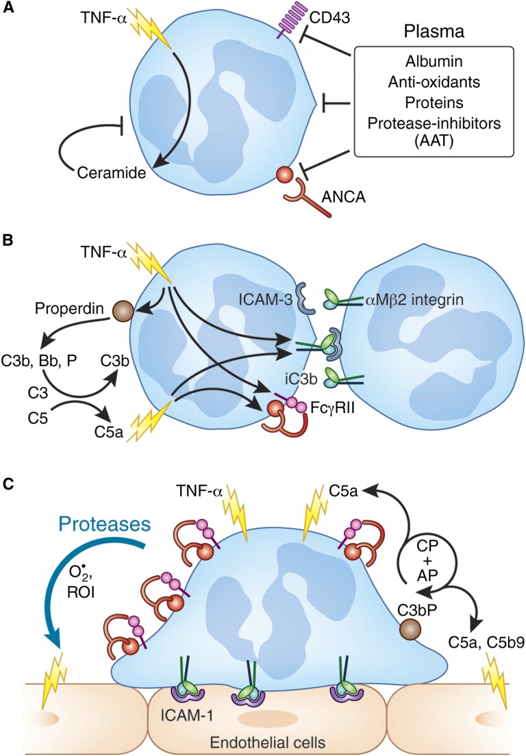
The two salient features of ANCA-associated vasculitis (AAV) are the restricted microvessel localization and the mechanism of inflammatory damage, independent of vascular immune deposits. The microvessel localization of the disease is due to the ANCA antigen accessibility, which is restricted to the membrane of neutrophils engaged in β2-integrin-mediated adhesion, while these antigens are cytoplasmic and inaccessible in resting neutrophils. The inflammatory vascular damage is the consequence of maximal proinflammatory responses of neutrophils, which face cumulative stimulations by TNF-α, β2-integrin engagement, C5a, and ANCA by the FcγRII receptor. This results in the premature intravascular explosive release by adherent neutrophils of all of their available weapons, normally designed to kill IgG-opsonized bacteria after migration in infected tissues.


Figures

Figure 1.
Figure 1.
Neutrophil-endothelial cell interactions. The classic view of neutrophil interactions with activated endothelium is a three-step process, although the development of intravital imaging recently revealed intermediate steps, such as the slow rolling and the intravascular crawling. Step 1 entails tethering and rolling involving selectins. Proinflammatory cytokines (TNF-α, IL1-β) or LPS induce the expression of endothelial selectins, able to interact with different ligands (PSGL-1, ESL-1, CD44) on neutrophils. Selectin engagement mediates rolling and, together with chemokines induced on the endothelial surface, initiates the “inside-out” activation of neutrophil β2-integrins. The interaction of partly activated β2-integrins leads first to neutrophils slow rolling on endothelial ICAM-1 and E-selectin. During this slow rolling, leukocytes integrate signals from chemokines or lipid mediators important for downstream events. Step 2 entails firm attachment via integrins. The level of intracellular calcium rises, leading to full β2-integrin activation and firm arrest on ICAMs. Chemokines trigger the polarization of leukocytes, with the formation of leading and trailing edges. The activation of αMβ2 (Mac-1) at the leading edge promotes intravascular crawling on the luminal surface to the point of transmigration. Intravital imaging allows for distinguishing two ways of transendothelial migration. As shown in step 3, the paracellular migration through endothelial junctions involves homotypic PECAM-1 and junctional adhesion molecule-A (JAM) interactions, resulting in unzipping of the endothelial cell junctions. In the transcellular migration, high density of ICAM-1 and VCAM-1 on specialized docking structures (migratory cup) captures crawling neutrophils and facilitates their way through the endothelial cells.,
Figure 2.
Figure 2.
Neutrophil activation promoted by TNF-α and amplified by ANCA. (A) Homeostatic control of TNF-induced neutrophil activation in the blood flow. Plasma proteins and oxidants prevent untimely intravascular activation of neutrophils. In particular, serum albumin prevents the shedding of leukosialin (CD43), a prerequisite for neutrophil adhesion and inhibits most neutrophil responses to low concentration of inflammatory stimuli. Neutrophil degranulation and oxidative responses are delayed by the endogenous ceramide, generated upon TNF-α priming. Finally, anti-PR3 ANCA do not react with membrane PR3 in the presence of plasma and particularly of α1-antitrypsin. (B) Initial step of low-grade activation of circulating neutrophils involving cell-cell homotypic interactions. TNF-stimulated neutrophils activate the complement alternative pathway via the secretion of endogenous properdin. This releases the powerful agonist C5a, able to promote homotypic aggregation of circulating neutrophils, via interactions of the TNF-activated αMβ2 (Mac-1)-integrins with ICAM-3 or iC3b on bystander neutrophils. TNF/β2-integrin joint signals would promote sufficient ANCA antigen membrane expression to overcome the plasma inhibitory effect and allow the access of ANCA Fab to their antigen, whereas their Fc portion interacts with FcγRIIa receptors. By doing so, ANCA give the final signals for the firm adhesion of neutrophils to the endothelium. (C) Explosive responses of neutrophils adherent to endothelial cells in the presence of ANCA. Neutrophils then adhere to ICAM-1–expressing endothelial cells. During this firm adhesion, TNF/β2-integrin joint signals result in degranulation and oxidative burst, and a massive increase of ANCA antigen expression, particularly mPR3, on the neutrophil surface. Neutrophil-bound ANCAs trigger the complement classic pathway (CP), exclusively on adherent neutrophils and in synergy with the alternative pathway (AP) activated on TNF-stimulated cells. C5a fragments and C5b-9 soluble complexes, released close to endothelial cells, are able to activate these cells and to promote cell retraction and endothelium permeability.,, The synergy between signals promoted by TNF, β2-integrin engagement, and ANCA-bound FcγR leads to an intense degranulation and the release of proteases and oxidants highly toxic for the endothelium.
Figure 3.
Figure 3.
Clustering, in lipid rafts, of neutrophil functional receptors required for ANCA-mediated responses. The ANCA-induced oxidative burst is promoted by joint signals triggered by TNF priming, β2-integrin engagement, and the clustering of Fcγ-receptors.,,, TNF-α mobilizes intracellular pools of αMβ2-integrin (Mac-1), nicotinamide adenine dinucleotide phosphate (NADPH) oxidase protein gp91phox, and PR3 from secretory vesicles to the plasma membrane, resulting in a co-localization of gp91phox, β2-integrins, and FcγRII. TNF-α, together with the engagement of β2-integrins, triggers the clustering of FcγRII receptors required for FcγR-induced signaling. The same signals induce the association of β2-integrins, NADPH-oxidase components, and FcγRII with the cytoskeleton, the latter being increased in the presence of anti-PR3 or anti-MPO antibodies., Upon TNF-α priming, PR3 forms a complex with NB1 and with CD11b/CD18, all three proteins being localized in cholesterol-rich domains (lipid rafts),, together with signaling molecules, cross-linked FcγRII,, and NADPH-oxidase membrane components.
Figure 4.
Figure 4.
Renal small vessels. AAV results in necrotic and crescentic GN and lung hemorrhage and is characterized by microvascular inflammation and necrosis in a variety of organs. The two organs that are most extensively injured in this manner are the kidney, through the development of pauci-immune crescentic GN, and the lung, with consequent alveolar hemorrhage. In the kidney, AAV preferentially involves glomerular and peritubular capillaries and venules, and arterioles. AAV does not affect interlobar and arcuate arteries and rarely interlobular arteries. Thus, the vascular distributions of AAV and of medium-sized-vessel and large-vessel vasculitides minimally overlap each other. The predilection of AAV for glomerular capillaries (necrotic capillaritis in A) and peritubular capillaries and venules (peritubular inflammatory capillaritis in B) is so exclusive that it is likely to be related the unique structure of renal capillaries. Interestingly, peritubular inflammatory capillaritis may be observed in AAV with few, and even without any, glomerular necrosis, suggesting that distinct mechanism of neutrophil-endothelial cell interactions are involved in AAV in glomeruli and in peritubular capillaries.
Figure 5.
Figure 5.
Nonconventional pathways of leukocyte recruitment in glomeruli. (A) Intravascular physical trapping independently of endothelial adhesion molecules. Neutrophil sequestration in capillaries results from ANCA-induced actin polymerization (green line) and increased cell rigidity and homotypic aggregation, mediated by TNF-activated β2-integrins interacting with ICAM-3 on bystander neutrophils. (B) Glomerular leukocyte adhesion induced by anti-GBM in mice and involving platelets. After anti-GBM antibody or immune complex deposition, platelets adhere to the glomerular endothelial lesions via platelet glycoprotein VI, fibrinogen being a potential ligand. Activated platelet express P-selectin, which recruits neutrophils by binding PSGL-1. The resulting neutrophil adhesion, which also involves β2-integrin/ICAM-1 interactions but bypasses the initial requirement for rolling, is restricted to glomerular capillaries and not observed in postcapillary venules. (C) Glomerular leukocyte adhesion induced by high doses of anti-MPO in mice. Infusion of high doses of anti-MPO antibody to LPS-free mice resulted in a glomerular leukocyte adhesion, which was β2-integrin independent, but instead required neutrophil α4-integrin and an unknown ligand distinct from VCAM-1. Human neutrophils do not express α4β1, except in extreme septic conditions, but they express the α9β1-integrin, which has similar specificities., (D) Anti-MPO–induced glomerular leukocyte adhesion in LPS-treated mice. In this AAV experimental model, the synergistic effect of LPS with anti-MPO antibodies is directly related to both LPS-induced TNF synthesis and to increased expressions of CXCL1, CXCL2 (homologs of human IL-8), which participate in neutrophil glomerular recruitment. TNF-primed neutrophils, bearing anti-MPO antibodies, adhere to the endothelium of glomerular capillaries, via the β2-integrin LFA-1 interacting with endothelial ICAM-1.
Figure 6.
Figure 6.
Consequences of intravascular activation of adherent neutrophils by ANCA. (A) Endothelial cell (EC) lesions. During neutrophil adhesion to endothelium, β2-integrins induce the clustering of ICAM-1 molecules, which results in intraendothelial signaling and microvascular hyperpermeability,, by altering adherens junctions, and by increasing the caveolae-mediated transcytosis. Vascular permeability is enhanced by neutrophil proteases and oxidants and leukotrienes, such as LTB4, which play an important role, in particular via the release of neutrophil azurocidin (HBP). Neutrophil-secreted proteases (elastase, PR3) or oxidants also induce the apoptosis and the detachment of endothelial cells. Hyperpermeability results in plasma leakage and edema, whereas cell detachment results in the exposure of the prothrombotic subendothelial matrix. (B) Innate/adaptive immunity. Activated neutrophils participate in the recruitment, activation, and programming of antigen-presenting cells. Cathepsin G activates prochemerin to produce chemerin, one of the few cytokines that attracts both immature dendritic cells and plasmacytoid dendritic cells. Moreover, neutrophils secrete TNF-related ligand B-lymphocyte stimulator (BLyS), which helps to drive proliferation and maturation of B cell differentiation, and IFN-γ, which helps to drive differentiation of T cells and activation of macrophages. (C) NETosis. ANCA binding to TNF-primed neutrophils results in the release of extracellular traps NETs, similar to those observed after a microbial challenge. This neutrophil cell death mechanism, involving the nicotinamide adenine dinucleotide phosphate oxidase and the production of reactive oxygen species (ROS), produces webs of DNA covered with antimicrobial molecules, including the autoantigens PR3 and MPO and LL37. LL37 is able to convert the self DNA in an activator of plasmacytoid dendritic cells and may play a role in the transfer to adaptive immunity. Finally, ANCA from AAV patients binds to MPO and PR3 on NETs, indicating that epitopes of target autoantigens are present, which suggests that NETs might participate in the autoimmune response by providing high concentrations of extracellular autoantigens.

References

    1. Wainwright J, Davson J: The renal appearances in the microscopic form of periarteritis nodosa. J Pathol Bacteriol 62: 189–196, 1950 - PubMed
    1. Jennette JC, Falk RJ, Andrassy K, Bacon PA, Churg J, Gross WL, Hagen EC, Hoffman GS, Hunder GG, Kallenberg CG, McCluskey RT, Sinico RA, Rees AJ, van Es LA, Waldherr R, Wiik A: Nomenclature of systemic vasculitides. Proposal of an international consensus conference. Arthritis Rheum 37: 187–192, 1994 - PubMed
    1. Jennette JC, Falk RJ: Small-vessel vasculitis. N Engl J Med 337: 1512–1523, 1997 - PubMed
    1. van der Woude FJ, Rasmussen N, Lobatto S, Wiik A, Permin H, van Es LA, van der Giessen M, van der Hem GK, The TH: Autoantibodies against neutrophils and monocytes: Tool for diagnosis and marker of disease activity in Wegener’s granulomatosis. Lancet 1: 425–429, 1985 - PubMed
    1. Falk RJ, Jennette JC: Anti-neutrophil cytoplasmic autoantibodies with specificity for myeloperoxidase in patients with systemic vasculitis and idiopathic necrotizing and crescentic glomerulonephritis. N Engl J Med 318: 1651–1657, 1988 - PubMed

MeSH terms

Substances

LinkOut - more resources