. 2012 Oct 1;189(7):3767-76.
doi: 10.4049/jimmunol.1201216. Epub 2012 Aug 31.

Inducible tertiary lymphoid structures, autoimmunity, and exocrine dysfunction in a novel model of salivary gland inflammation in C57BL/6 mice

Affiliations

Inducible tertiary lymphoid structures, autoimmunity, and exocrine dysfunction in a novel model of salivary gland inflammation in C57BL/6 mice

Michele Bombardieri et al. J Immunol. .

Abstract

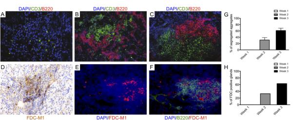
Salivary glands in patients with Sjögren's syndrome (SS) develop ectopic lymphoid structures (ELS) characterized by B/T cell compartmentalization, the formation of high endothelial venules, follicular dendritic cell networks, functional B cell activation with expression of activation-induced cytidine deaminase, as well as local differentiation of autoreactive plasma cells. The mechanisms that trigger ELS formation, autoimmunity, and exocrine dysfunction in SS are largely unknown. In this article, we present a novel model of inducible ectopic lymphoid tissue formation, breach of humoral self-tolerance, and salivary hypofunction after delivery of a replication-deficient adenovirus-5 in submandibular glands of C57BL/6 mice through retrograde excretory duct cannulation. In this model, inflammation rapidly and consistently evolves from diffuse infiltration toward the development of SS-like periductal lymphoid aggregates within 2 wk from AdV delivery. These infiltrates progressively acquire ELS features and support functional GL7(+)/activation-induced cytidine deaminase(+) germinal centers. Formation of ELS is preceded by ectopic expression of lymphoid chemokines CXCL13, CCL19, and lymphotoxin-β, and is associated with development of anti-nuclear Abs in up to 75% of mice. Finally, reduction in salivary flow was observed over 3 wk post-AdV infection, consistent with exocrine gland dysfunction as a consequence of the inflammatory response. This novel model has the potential to unravel the cellular and molecular mechanisms that regulate ELS formation and their role in exocrine dysfunction and autoimmunity in SS.


Figures

Figure 1
Figure 1. Efficiency of transfection following AdV delivery via retrograde mouse submandibular gland excretory duct cannulation
A-B) A glass cannula is inserted in the excretory orifice of the submandibular gland of a 12 weeks-old C57BL/6 mouse. C) Representative picture of a cannulated NOD mouse demonstrating that delivery of 50μl of bromophenol blue dye is sufficient to permeate the cannulated submandibular gland with no spillage to the contralateral gland or the adjacent sublingual gland (arrow). D) Representative dose-response time-course experiments of luciferase activity following delivery of LucAdV at 10^6, 10^7 and 10^8pfu/gland and vehicle control showing dose-dependent enzymatic activity within the first week. Dose dependency is lost at week 2 and 3 despite persistent transgene expression.
Figure 2
Figure 2. AdV delivery and its persistence in ductal epithelial cells progressively induce SS-like periductal inflammatory foci in wild-type C57BL/6 mice
A-F) Representative microphotographs showing inflammatory responses induced by local administration of either vehicle (A-C), LucAdV (D-F) or LacZAdV (G-I). After AdV delivery, diffuse lymphomonocytic infiltration in the first week (D, G) is followed by progressive organization of periductal inflammatory aggregates during week 2 (E, H) and week 3 pc (F, I). Vehicle delivery induces negligible inflammation (A-C). The mean ± SEM of the number of periductal foci/gland following either LucAdV or LacZAdV delivery at different time points is reported in J. (**p<0.01 between AdV and vehicle-treated mice at each time point, minimum of 10 mice per time point). G-I) Representative microphotographs of submandibular glands infected with LacZAdV and stained for beta-galactosidase (XGal, with nuclear counterstaining with haematoxylin). Numerous beta-galactosidase-positive cells are present in the gland within the first week pc (G, arrowheads). By week 2 pc only ductal epithelial cells, often surrounded by periductal inflammatory infiltrates, show enzymatic activity (H). By week 3 pc no residual XGal-positive cells are detectable (I) but high sensitivity luciferase activity assay in LucAdV-infected glands shows persistent signal well above background levels up to three weeks pc (K, mean ± SEM of luciferase activity. Original magnification in A-I x100. (***p<0.001 between AdV-treated and vehicle-treated mice at each time point, minimum of 10 mice per time point).
Figure 3
Figure 3. Progressive development of T/B cell segregation and FDC networks following AdV infection in the submandibular glands of C57BL/6 mice
A-C) Microphotographs showing double immunofluorescent staining for CD3 (T cells, in green) and B220 (B cells, in red) of frozen sections from AdV-treated C57BL/6 salivary glands. Nuclei are counterstained with DAPI (blue). Early diffuse infiltration is dominated by T cells (A) with subsequent recruitment of B cells showing initial T/B cell compartmentalization within the first 2 weeks pc (B). By week 3 pc over 60% of periductal foci display full segregation of T and B cells in separate areas (C and G). D-F) Microphotographs of AdV-treated C57BL/6 submandibular glands at week 3 pc showing differentiation of FDC networks as demonstrated by staining for FDC-M1 (D, in brown and E in red). FDC networks invariably develop in the context of the B cell rich area of the aggregates (F) in over 60% of AdV-treated C57BL/6 mice (H). Original magnification x200.
Figure 4
Figure 4. Development of ELS in C57BL/6 submandibular glands is preceded by ectopic expression of the lymphoid chemokines/Ltβ pathway
A-F) Quantitative TaqMan real-time PCR time-course analysis of CCL19 (and its receptor CCR7, A,D) CXCL13 (and its receptor CXCR5, B,E), and Ltβ(and its receptor, C,F) mRNA transcripts showing abundant up-regulation following AdV-delivery in the submandibular glands. Earliest upregulation was observed for CXCL13 and Ltβ mRNA while peak of expression of these factors was detectable in parallel with full histological development of ELS (A-E). Conversely, LtβR expression appears unaffected by AdV treatment (F). Data are expressed as mean ± SEM of the fold-increase compared to an internal calibrator. (*p<0.05, **p<0.01 and ***p<0.001 between AdV-treated and vehicle-treated mice at each time point, minimum of 10 mice per time point). G-N) Microphotographs of multi-color confocal microscopy analysis confirming protein expression of lymphoid chemokines CXCL13 (I, in blue) and CCL21 (M) and showing co-localization of CXCL13 within the B cell-rich area (G-J) and of CCL21 within the T cell rich area of the aggregate (K-N) B cells are shown in red (CD19) and T cells in green (CD3), respectively. Original magnification x200.
Figure 5
Figure 5. AdV-induced ELS in C57BL/6 submandibular glands acquire characteristics of functional ectopic germinal centers
A-D) Sequential immunofluorescence staining of AdV-treated submandibular gland sections of C57BL/6 mice at 3 weeks pc showing that highly organized ELS characterized by high-endothelial venules (HEV, A in red), T/B cell segregation (B, B220 in red and CD3 in green) and FDC networks (C) support the differentiation of GL7+ germinal center B cells (D, B220 in red and GL7 in green). E-H) Quantitative TaqMan real-time PCR time-course analysis of salivary gland expression of AID mRNA (E) and transcripts for cytokines downstream AID activation, such as IL-4, BAFF and IL-21 (F-H). AID expression peaks in parallel with the development of fully formed ELS and is associated with significant upregulation of IL-4, BAFF and IL-21. Data are expressed as mean ± SEM of the fold-increase compared to an internal calibrator. (*p<0.05 and **p<0.01 between AdV-treated and vehicle-treated mice at each time point, minimum of 10 mice per time point). Original magnification X200 (A-D).
Figure 6
Figure 6. Development of anti-viral and anti-nuclear antibodies following AdV delivery in the submandibular glands of C57BL/6 mice
A) Western blot showing progressive development of anti-AdV IgG in the serum of AdV-treated but not vehicle-cannulated mice. Protein extracts from AdV-infected (+) and uninfected (−) 293 cells were run in parallel. From left to right: vehicle-treated C57BL/6 mouse serum at 20 days pc, commercial anti-AdV antiserum, sera from AdV-cannulated mice at different time-points (5, 9, 12, 15, 20, 23 days pc). Anti-AdV responses are observed as early as day 5 pc and are mainly directed against proteins of the viral core and capside. B-E) Representative microphotographs of immunofluorescent detection of anti-nuclear antibodies (ANA) using Hep2 cells as substrate (B,C) demonstrating strong nuclear reactivity in sera of AdV-treated (C) but not in vehicle-cannulated (B) C57BL/6 mice 3 weeks pc (dilution 1:80). ANA reactivity was progressively evident over time in AdV-cannulatedC57BL/6 mice (D) with 75% of mice displaying ANA positivity by week 3 pc (E). Original magnification X400 (B-C).
Figure 7
Figure 7. Sustained reduction in salivary flow following AdV infection of C57BL/6 mice submandibular glands
Column graphs comparing salivary flow in the AdV-treated submandibular and the contralateral gland at week 1, 2 and 3 pc. Salivary flow is measured as mg of saliva produced in 10 minutes/body weight following pilocarpine stimulation (see methods). Exocrine dysfunction in the AdV-cannulated glands can be observed up to three weeks pc. Data are expressed as mean ± SEM. (*p<0.05 between AdV-treated and vehicle-treated mice at each time point, minimum of 5 mice per time point).

Similar articles

Cited by

References

    1. Drayton DL, Liao S, Mounzer RH, Ruddle NH. Lymphoid organ development: from ontogeny to neogenesis. Nat Immunol. 2006;7:344–353. - PubMed
    1. Aloisi F, Pujol-Borrell R. Lymphoid neogenesis in chronic inflammatory diseases. Nat Rev Immunol. 2006;6:205–217. - PubMed
    1. Manzo A, Bombardieri M, Humby F, Pitzalis C. Secondary and ectopic lymphoid tissue responses in rheumatoid arthritis: from inflammation to autoimmunity and tissue damage/remodeling. Immunol Rev. 2010;233:267–285. - PubMed
    1. Routsias JG, Tzioufas AG, Moutsopoulos HM. The clinical value of intracellular autoantigens B-cell epitopes in systemic rheumatic diseases. Clin Chim Acta. 2004;340:1–25. - PubMed
    1. Chisholm DM, Mason DK. Labial salivary gland biopsy in Sjogren’s disease. J Clin Pathol. 1968;21:656–660. - PMC - PubMed

Publication types