. 2012 Oct 1;189(7):3585-92.
doi: 10.4049/jimmunol.1102408. Epub 2012 Aug 31.

Cyanovirin-N inhibits mannose-dependent Mycobacterium-C-type lectin interactions but does not protect against murine tuberculosis

Affiliations

Cyanovirin-N inhibits mannose-dependent Mycobacterium-C-type lectin interactions but does not protect against murine tuberculosis

Nicole N Driessen et al. J Immunol. .

Abstract

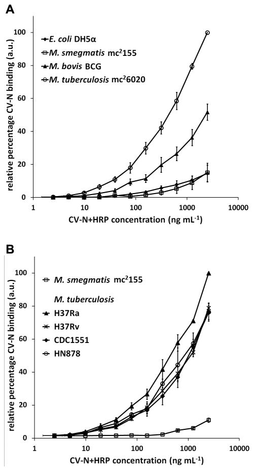
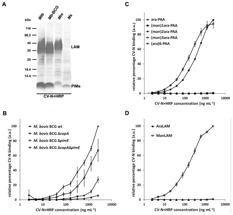
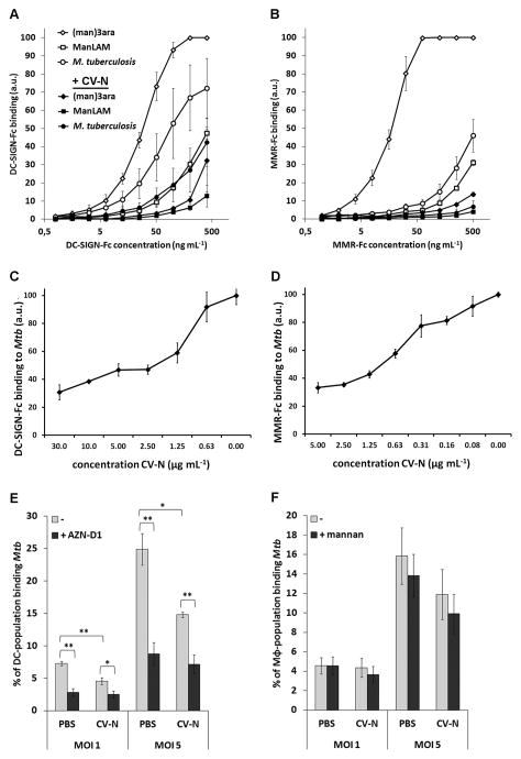
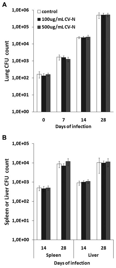
Cyanovirin-N (CV-N) is a mannose-binding lectin that inhibits HIV-1 infection by blocking mannose-dependent target cell entry via C-type lectins. Like HIV-1, Mycobacterium tuberculosis expresses mannosylated surface structures and exploits C-type lectins to gain cell access. In this study, we investigated whether CV-N, like HIV-1, can inhibit M. tuberculosis infection. We found that CV-N specifically interacted with mycobacteria by binding to the mannose-capped lipoglycan lipoarabinomannan. Furthermore, CV-N competed with the C-type lectins DC-SIGN and mannose receptor for ligand binding and inhibited the binding of M. tuberculosis to dendritic cells but, unexpectedly, not to macrophages. Subsequent in vivo infection experiments in a mouse model demonstrated that, despite its activity, CV-N did not inhibit or delay M. tuberculosis infection. This outcome argues against a critical role for mannose-dependent C-type lectin interactions during the initial stages of murine M. tuberculosis infection and suggests that, depending on the circumstances, M. tuberculosis can productively infect cells using different modes of entry.


Conflict of interest statement

Conflict of interest

The authors declare that no conflict of interest exists related to this work.

Figures

FIGURE 1
FIGURE 1
CV-N differentially recognizes various mycobacterial species. A, M. tuberculosis strain mc26020, M. bovis BCG strain Copenhagen, M. smegmatis strain mc2155, and E. coli strain DH5α were coated on a 96-well plate and probed with a serial dilution of CV-N+HRP. CV-N binding was detected using OPD as coloring reagent and absorption was measured at 490 nm. B, In a similar assay, binding of CV-N to M. tuberculosis strains H37Rv, H37Ra, CDC1551, and HN878 was tested. In all cases, the data represent the average of three independent experiments plus the standard error of mean.
FIGURE 2
FIGURE 2
CV-N binds the α(1,2)-linked mannosyl residues present in the mannose caps of ManLAM and in PIM6. A, Cell lysates of M. tuberculosis mc26020 (Mtb), M. bovis BCG (Mb-BCG), M. marinum E11 (Mm), and M. smegmatis mc2155 (Ms) were examined on SDS-PAGE/immunoblot with CV-N+HRP. CV-N+HRP binds to ManLAM of M. tuberculosis, M. bovis BCG, and M. marinum, but not to PILAM of M. smegmatis. Staining with CV-N+HRP is also seen at the position of PIMs on the immunoblot. See Supplemental Fig. 1A for staining of the same cell lysates on immunoblot with (Man)LAM- and PIM6-specific antibodies. B, Whole cells of M. bovis BCG wild-type, ΔcapA (deficient for the mannose cap on LAM), ΔpimE (deficient in biosynthesis of PIM6), and double knockout ΔcapAΔpimE were examined for CV-N+HRP binding in whole-cell ELISA. The average of three independent experiments is shown. See Supplemental Fig. 1B for concomitant SDS-PAGE/immunoblot with M. bovis BCG cell lysates. C, PAA-coupled glycoconjugates and, D, lipoglycans ManLAM and AraLAM were coated on a 96-well plate and probed with a serial dilution of CV-N+HRP. The average of three independent experiments is shown plus the standard error of mean. Note, in C, binding of CV-N+HRP to glycoconjugates (ara)6-PAA, ara-PAA and (man)1ara-PAA, was below detection levels (i.e. 0.0 in figure) in all three cases.
FIGURE 3
FIGURE 3
CV-N inhibits binding of M. tuberculosis mc26020 or mycobacterial mannosylated structures to DC-SIGN-Fc, MMR-Fc, and to MoDCs, but not to MoMϕ. AB, Whole M. tuberculosis mc26020 cells, (man)3ara-PAA, and ManLAM were coated on a 96-well plate and incubated with serial dilutions of either A, DC-SIGN-Fc (starting at 400 ng mL−1), or B, MMR-Fc (starting at 500 ng mL−1) in the presence or absence of 50 μg mL−1 CV-N. The average of three independent experiments is shown plus the standard error of mean. CD, CV-N inhibits binding of M. tuberculosis mc26020 by C, DC-SIGN-Fc and D, MMR-Fc in a dose-dependent manner. Whole M. tuberculosis mc26020 cells were coated on a 96-well plate and incubated DC-SIGN-Fc (400 ng mL−1), or MMR-Fc (500 ng mL−1) in the absence or presence of a dilution series of CV-N. Shown is one representative experiment with average of triplicates plus standard deviation. E–F, M. tuberculosis cells were fluorescently labeled, diluted to the correct MOI, pre-treated with PBS or 200 μg mL−1 CV-N, washed, and added to E, MoDCs and, F, MoMϕ. MoDCs and MoMϕ were used untreated, or after pre-incubation with the DC-SIGN-specific inhibitory antibody AZN-D1 or with 2 mg mL−1 mannan, respectively. The percentage of the cell population binding M. tuberculosis was determined by flow cytometry. Shown is the average of results from five donors plus the standard error of mean. * p < 0.05; ** p < 0.005.
FIGURE 4
FIGURE 4
Aerosolic infection of mice with untreated or CV-N-pre-treated M. tuberculosis H37Rv. Mycobacteria were pre-incubated with PBS (‘untreated’) or with CV-N at concentrations of either 100 μg mL−1 or 500 μg mL−1 (is comparable CV-N:bacteria ratio as used for pre-treatment of the MOI5-suspensions in the in vitro assays in Figure 3EF, or five-fold higher, respectively). Mice were infected with the untreated or CV-N-treated M. tuberculosis via aerosolic solution on day 0. CFUs were counted in A, lung, and B, spleen and liver. The CFU count on day 0 and day 7 for spleen and liver was zero in all mice. Shown is median of five mice per group plus median absolute deviation.

References

    1. World Health Organization. Global tuberculosis control: WHO report 2010. WHO Press; Switzerland: 2010. Report No.: WHO/HTM/TB/2010.7.
    1. Fine PE. Variation in protection by BCG: implications of and for heterologous immunity. Lancet. 1995;346:1339–1345. - PubMed
    1. Torrelles JB, Schlesinger LS. Diversity in Mycobacterium tuberculosis mannosylated cell wall determinants impacts adaptation to the host. Tuberculosis (Edinb) 2010;90:84–93. - PMC - PubMed
    1. Schlesinger LS. Macrophage phagocytosis of virulent but not attenuated strains of Mycobacterium tuberculosis is mediated by mannose receptors in addition to complement receptors. J Immunol. 1993;150:2920–2930. - PubMed
    1. Tailleux L, Schwartz O, Herrmann JL, Pivert E, Jackson M, Amara A, Legres L, Dreher D, Nicod LP, Gluckman JC, Lagrange PH, Gicquel B, Neyrolles O. DC-SIGN is the major Mycobacterium tuberculosis receptor on human dendritic cells. J Exp Med. 2003;197:121–127. - PMC - PubMed

Publication types

MeSH terms