

Balloon-occluded Retrograde Transvenous Obliteration (BRTO): Preprocedural Evaluation and Imaging
- PMID: 22942546
- PMCID: PMC3312157
- DOI: 10.1055/s-0031-1284455
Balloon-occluded Retrograde Transvenous Obliteration (BRTO): Preprocedural Evaluation and Imaging
Abstract
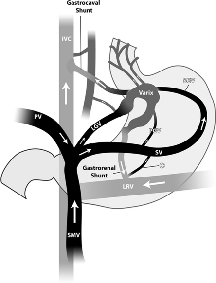
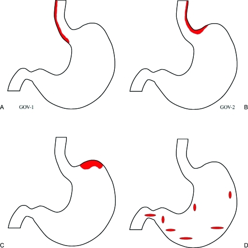
Patients undergoing balloon retrograde transvenous obliteration (BRTO) are mostly decompensated cirrhotic with either bleeding gastric varices (GV) or hepatic encephalopathy. It is crucial that clinicians are up-to-date with the assessments needed prior to BRTO to anticipate and prevent complications, and to deliver critical quality care. These patients will require preprocedural assessments and management, including endoscopic, clinical, laboratory, and imaging evaluation. Endoscopic evaluation is mandatory prior to BRTO, and it is highly recommended that it be performed at the same institution where BRTO will be performed. It is essential that clinicians are aware of the potential benefits and complications that may result from BRTO. These complications should be anticipated and prevented when possible. For GV bleeders, there should be consideration of a transvenous intrahepatic portosystemic shunt (TIPS) during or before BRTO in patients with refractory ascites or pleural effusion, as well as endoscopic banding or a TIPS in patients with high-risk esophageal varices. Patients undergoing BRTO are usually complicated and require a team approach. In this article, the authors address these assessment and preparatory management and planning procedures prior to the BRTO procedure as well as expected outcomes and potential complications.
Keywords: BRTO; Gastric varices; TIPS; liver cirrhosis; portal hypertension; splenorenal shunt.
Figures
References
-
- Kanagawa H, Mima S, Kouyama H, Gotoh K, Uchida T, Okuda K. Treatment of gastric fundal varices by balloon-occluded retrograde transvenous obliteration. J Gastroenterol Hepatol. 1996;11(1):51–58. - PubMed
-
- Koito K, Namieno T, Nagakawa T, Morita K. Balloon-occluded retrograde transvenous obliteration for gastric varices with gastrorenal or gastrocaval collaterals. AJR Am J Roentgenol. 1996;167(5):1317–1320. - PubMed
-
- Kitamoto M, Imamura M, Kamada K, et al. Balloon-occluded retrograde transvenous obliteration of gastric fundal varices with hemorrhage. AJR Am J Roentgenol. 2002;178(5):1167–1174. - PubMed
-
- Sakurabayashi S, Sezai S, Yamamoto Y, Hirano M, Oka H. Embolization of portal-systemic shunts in cirrhotic patients with chronic recurrent hepatic encephalopathy. Cardiovasc Intervent Radiol. 1997;20(2):120–124. - PubMed
-
- Fukuda T, Hirota S, Sugimura K. Long-term results of balloon-occluded retrograde transvenous obliteration for the treatment of gastric varices and hepatic encephalopathy. J Vasc Interv Radiol. 2001;12(3):327–336. - PubMed
