. 2012;13(7):8126-8141.
doi: 10.3390/ijms13078126. Epub 2012 Jun 29.

Elongation factor 1β' gene from Spodoptera exigua: characterization and function identification through RNA interference

Affiliations

Elongation factor 1β' gene from Spodoptera exigua: characterization and function identification through RNA interference

Li-Na Zhao et al. Int J Mol Sci. 2012.

Abstract

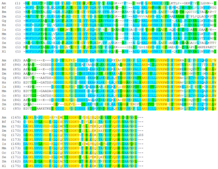
Elongation factor (EF) is a key regulation factor for translation in many organisms, including plants, bacteria, fungi, animals and insects. To investigate the nature and function of elongation factor 1β' from Spodoptera exigua (SeEF-1β'), its cDNA was cloned. This contained an open reading frame of 672 nucleotides encoding a protein of 223 amino acids with a predicted molecular weight of 24.04 kDa and pI of 4.53. Northern blotting revealed that SeEF-1β' mRNA is expressed in brain, epidermis, fat body, midgut, Malpighian tubules, ovary and tracheae. RT-PCR revealed that SeEF-1β' mRNA is expressed at different levels in fat body and whole body during different developmental stages. In RNAi experiments, the survival rate of insects injected with SeEF-1β' dsRNA was 58.7% at 36 h after injection, which was significantly lower than three control groups. Other elongation factors and transcription factors were also influenced when EF-1β' was suppressed. The results demonstrate that SeEF-1β' is a key gene in transcription in S. exigua.

Keywords: RNAi; Spodoptera exigua; cloning; elongation factor; expression pattern.


Figures

Figure 1
Figure 1
Nucleotide and amino acid sequences of EF-1β′ from the beet armyworm S. exigua. The nucleotide sequence reported in this paper has been submitted to GenBank under accession no. EU258621. Italic and overstriking nucleotides are start and stop codons, respectively. The primers for dsSeEF-1β′ RNA synthesis and detection are marked by an underline and double underline, respectively. The termination signal AATAAA is boxed.
Figure 2
Figure 2
Phylogenetic analysis of SeEF-1β′ and EF-1β′s from other animal species. The phylogenetic tree was constructed based on the amino acid sequences of known EFs. Full length amino acid sequences were aligned with the Mega 3.1 program to generate the phylogenetic tree. A bootstrap analysis was carried out, and the robustness of each cluster was verified with 1000 replicates. Values at the cluster branches indicate the results of the bootstrap analysis. The scale on the x-axis represents the estimated branch lengths. EF-1β′s were from A. pisum (NM_001162346), A. aegypti (AY552052), A. gambiae (XM_558148), A. mellifera (XM_625024), B. mori (NM_001044091), B. floridae (XP_002227182), C. quinquefasciatus (XM_001847313), D. citri (DQ673433), D. melanogaster (NM_080069), E. lucius (BT079178), G. gallus (AJ721003), H. sapiens (CH236950), I. scapularis (DQ066216), M. hirsutus (EF070471), M. musculus (AK012756), N. vitripennis (XM_001599881), O. cuniculus (NM_001082399), O. mordax (BT074513), P. xylostella (AB180443), S. mansoni (FN357357), S. exigua (EU258621), T. infestans (EF639083), T. castaneum (XM_968676)and X. laevis (NM_001090665) (all sequences from GeneBank).
Figure 3
Figure 3
Alignment of EF-1β′ proteins from insects and other animals. EF-1β′s were from A. mellifera (Am), B. mori (Bm), B. floridae (Bf), D. citri (Dc), D. melanogaster (Dm), G. gallus (Gg), H. sapiens (Hs), I. scapularis (Is), M. musculus (Mm), O. cuniculus (Oc), S. mansoni (Sm), S. exigua (Se), and X. laevis (Xl). GenBank accession numbers (DNA) are as in Figure 2. Highly conserved regions are shown in yellow, green and sky-blue.
Figure 4
Figure 4
Northern blot analyses of the SeEF-1β′ transcript in different tissues of fifth instar S. exigua larvae. Total RNA was extracted from various tissues: Brain (Br), Epidermis (Ep), Fat body (Fb), Midgut (Mg), Malpighian tubules (Mt), Ovary (Ov) and Tracheae (Tr). An SeEF-1β′-specific probe was radiolabeled with (α-32P)-dCTP. Following hybridization and detection by autoradiography using the SeEF-1β′ probe, the membrane was stripped by boiling in 0.1% SDS. The rRNA was used as a reference.
Figure 5
Figure 5
Developmental expression of S. exigua EF-1β′ mRNA in the fat body (A) and whole body (B). [α-32P] dCTP labeled SeEF-1β′ cDNA, which was amplified using specific primers EF-FP and EF-RP, was used as a probe. β-actin was labeled with [α-32P] dCTP as a control. (A) RNA was extracted from the fat body every 24 h from fifth instar larvae (5L), wandering (pre-pupae) larvae (W) and pupae (P) 5L1-5L3 means the first to third day of the fifth instar larvae. W means pre-pupae larvae; P1-P7 means the first to seventh day of papae; (B) RNA was extracted from the whole body from first instar larvae to the fifth day of pupae (P). 1L1-5L4 means the first day of the first instar larvae to fourth day of the fifth instar larvae; W1 and W2 means the first and second day of pre-pupae larvae; P1-P5 means the first to fifth day of pupae. β-Actin was used as a loading control.
Figure 5
Figure 5
Developmental expression of S. exigua EF-1β′ mRNA in the fat body (A) and whole body (B). [α-32P] dCTP labeled SeEF-1β′ cDNA, which was amplified using specific primers EF-FP and EF-RP, was used as a probe. β-actin was labeled with [α-32P] dCTP as a control. (A) RNA was extracted from the fat body every 24 h from fifth instar larvae (5L), wandering (pre-pupae) larvae (W) and pupae (P) 5L1-5L3 means the first to third day of the fifth instar larvae. W means pre-pupae larvae; P1-P7 means the first to seventh day of papae; (B) RNA was extracted from the whole body from first instar larvae to the fifth day of pupae (P). 1L1-5L4 means the first day of the first instar larvae to fourth day of the fifth instar larvae; W1 and W2 means the first and second day of pre-pupae larvae; P1-P5 means the first to fifth day of pupae. β-Actin was used as a loading control.
Figure 6
Figure 6
S. exigua survival rates and RT-PCR analysis of SeEF-1β′ transcripts after injection of dsRNA. (A) Survival rates of insects at different times after injection of dsSeEF-1β′, dsGFP, DEPC water alone and no injection. Survival rates of insects based on key developmental stages for time intervals of 24 h, 36 h, 48 h (pre-pupae stage), 60 h (pupation stage) and 204 h (eclosion stage) post-injection. Percentage values were arcsine square root transformed prior to analyses to correct for the non-normal distribution of percentage values. Different letters in the same injection stage indicate significant difference of the survival rates (p, 0.05, Duncan’s test). No significant difference was found by ANOVA (p, 0.05). All error bars represent standard deviation (n = 3); (B) Three lively insects were obtained at 24 h, 36 h, 48 h, 72 h, 96 h, 120 h and 168 h after injection at random. Total RNA was extracted and SeEF-1β′ transcripts were detected using RT-PCR. The post-injection time for 24 h, 36 h, 48 h, 72 h, 96 h, 120 h and 168 h are marked. The rRNA and housekeeping gene of β-actin were used as references.
Figure 7
Figure 7
RT-PCR analysis of SeEF-1α, SeEF-2 and SeFH mRNA expression after injection of ds SeEF-1β′. Three lively insects were obtained at 12 h, 24 h, 36 h, 48 h, 72 h and 96 h after injection at rando. Total RNA was extracted and SeEF-1α, SeEF-2 and SeFH transcripts were detected using RT-PCR. The rRNA and housekeeping gene of β-actin were used as references. (A) SeEF-1α gene’s expression after injected dsSeEF-1β′, dsGFP and no injection; (B) SeEF-2 gene’s expression after injected dsSeEF-1β′, dsGFP and no injection; (C) SeFH gene’s expression after injected dsSeEF-1β′, dsGFP and no injection.

Similar articles

Cited by

References

    1. Linz J.E., Sypherd P.S. Expression of three genes for elongation factor 1α during morphogenesis of Mucor racemosus. Mol. Cell. Biol. 1987;7:1925–1932. - PMC - PubMed
    1. Riis B., Rattan S.I.S., Clark B.F.C., Merrick W.C. Eukaryotic protein elongation factors. Trends Biochem. Sci. 1990;15:420–424. - PubMed
    1. Bassel G.J., Powers C.M., Taneja K.L., Singer R.H. Single mRNAs visualized by ultrastructural in situ hybridization are principally localized at actin filament intersections in fibroblasts. J. Cell Biol. 1994;126:863–876. - PMC - PubMed
    1. Margutti P., Ortona E., Vaccari S., Barca S., Riganò R., Teggi A., Muhschlegel F., Frosch M., Siracusano A. Cloning and expression of a cDNA encoding an elongation factor 1β/δ protein from Echinococcus granulosus with immunogenic activity. Parasite Immunol. 1999;21:485–492. - PubMed
    1. Fujita T., Piuz I., Schlegel W. The transcription elongation factors NELF, DSIF and P-TEFb control constitutive transcription in a gene-specific manner. FEBS Lett. 2009;583:2893–2898. - PubMed

Publication types