. 2012;13(7):8834-8852.
doi: 10.3390/ijms13078834. Epub 2012 Jul 16.

The tricyclodecan-9-yl-xanthogenate D609 triggers ceramide increase and enhances FasL-induced caspase-dependent and -independent cell death in T lymphocytes

Affiliations

The tricyclodecan-9-yl-xanthogenate D609 triggers ceramide increase and enhances FasL-induced caspase-dependent and -independent cell death in T lymphocytes

Delphine Milhas et al. Int J Mol Sci. 2012.

Abstract

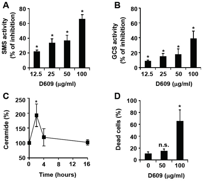
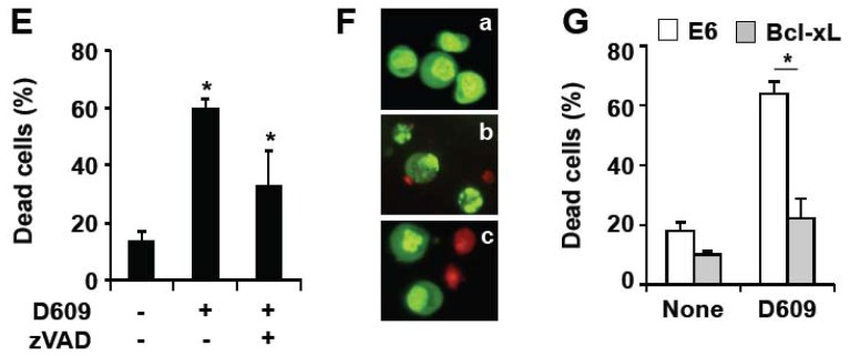
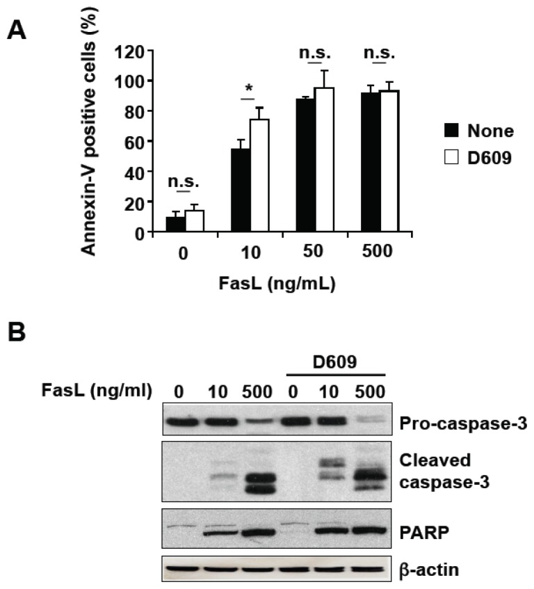
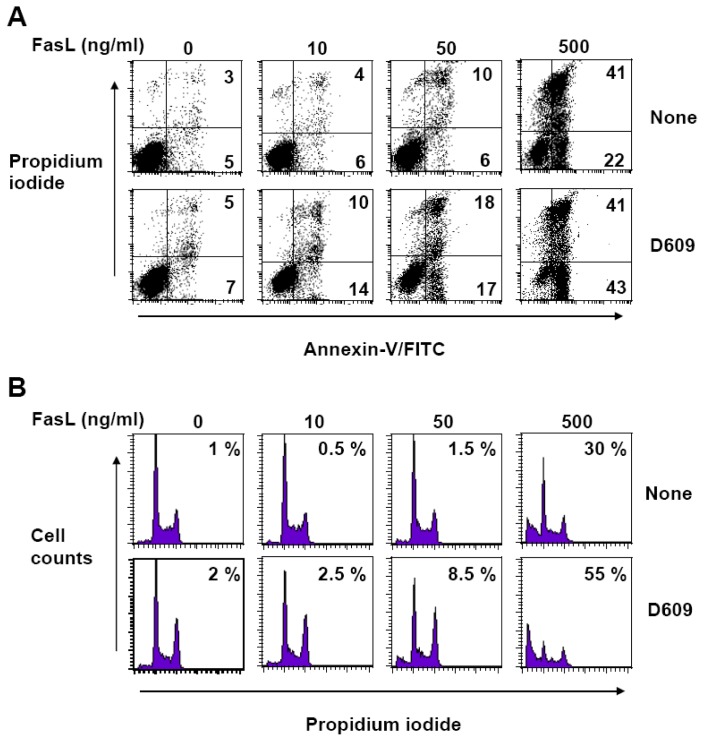
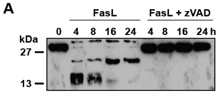
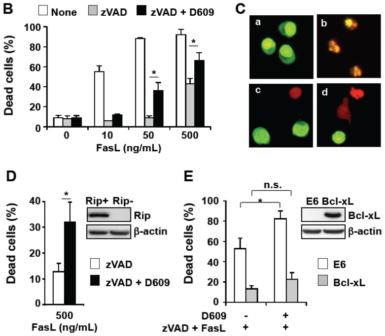
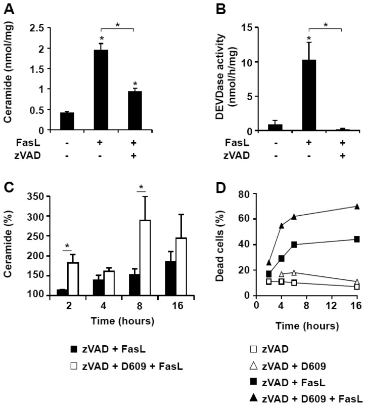
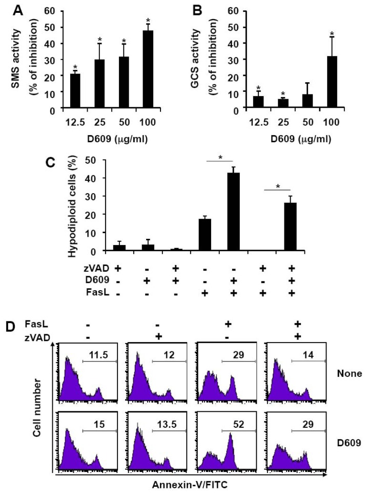
D609 is known to modulate death receptor-induced ceramide generation and cell death. We show that in Jurkat cells, non-toxic D609 concentrations inhibit sphingomyelin synthase and, to a lesser extent, glucosylceramide synthase, and transiently increase the intracellular ceramide level. D609 significantly enhanced FasL-induced caspase activation and apoptosis. D609 stimulated FasL-induced cell death in caspase-8-deficient Jurkat cells, indicating that D609 acts downstream of caspase-8. At high FasL concentration (500 ng/mL), cell death was significantly, but not completely, inhibited by zVAD-fmk, a broad-spectrum caspase inhibitor, indicating that FasL can activate both caspase-dependent and -independent cell death signaling pathways. FasL-induced caspase activation was abolished by zVAD-fmk, whereas ceramide production was only partially impaired. D609 enhanced caspase-independent ceramide increase and cell death in response to FasL. Also, D609 overcame zVAD-fmk-conferred resistance to a FasL concentration as low as 50 ng/mL and bypassed RIP deficiency. It is likely that mitochondrial events were involved, since Bcl-xL over-expression impaired D609 effects. In PHA-activated human T lymphocytes, D609 enhanced FasL-induced cell death in the presence or absence of zVAD-fmk. Altogether, our data strongly indicate that the inhibition of ceramide conversion to complex sphingolipids by D609 is accompanied by an enhancement of FasL-induced caspase-dependent and -independent cell death in T lymphocytes.

Keywords: ALPS; CD95; apoptosis; glucosylceramide synthase; necrosis; sphingomyelin synthase.


Figures

Figure 1
Figure 1
D609 inhibits SMS and GCS activities, and triggers ceramide increase and subsequent cell death in Jurkat cells. (A,B) Jurkat cells (clone A3) were incubated in the presence or absence of the indicated concentrations of D609 for 1 h and further incubated for 2 h in the presence of 2.5 μM C6-NBD-ceramide. SMS (A) and GCS (B) activities were determined by quantifying fluorescent SM and GlcCer. Results are expressed as the % of inhibition of the activities measured in the absence of D609. (C) A3 cells were incubated for the indicated times with 50 μg/mL D609 and ceramide content was quantified. (DG) Cells were incubated for 16 h in the presence or absence of D609 (100 μg/mL or the indicated concentrations) and cell death was evaluated by microscopy analysis (F) or quantified by flow cytometry after annexin-V-FITC and propidium iodide labeling (D,E,G). At this incubation time, most of the annexin-V positive cells were also stained by propidium iodide and considered as dead cells. (E) Cells were pre-incubated for 1 h in the presence of 40 μM zVAD-fmk and further incubated for 16 h with 100 μg/mL D609 as indicated. (F) A3 cells were incubated with control medium (a), containing 100 μg/mL D609 (b) or a combination of 40 μM zVAD-fmk and 100 μg/mL D609 (c). After 16 h, cells were stained with Syto-13 (green probe) and propidium iodide and analyzed under a fluorescent microscope. G, Mock-transfected E6 Jurkat cells (E6) and Bcl-xL-over-expressing E6 cells (Bcl-xL) were incubated in the presence (D609) or absence (None) of 100 μg/mL D609 for 16 h. (AE,G) Values are means ± S.E.M. of three independent experiments. (F) Pictures are representative of three independent experiments.
Figure 1
Figure 1
D609 inhibits SMS and GCS activities, and triggers ceramide increase and subsequent cell death in Jurkat cells. (A,B) Jurkat cells (clone A3) were incubated in the presence or absence of the indicated concentrations of D609 for 1 h and further incubated for 2 h in the presence of 2.5 μM C6-NBD-ceramide. SMS (A) and GCS (B) activities were determined by quantifying fluorescent SM and GlcCer. Results are expressed as the % of inhibition of the activities measured in the absence of D609. (C) A3 cells were incubated for the indicated times with 50 μg/mL D609 and ceramide content was quantified. (DG) Cells were incubated for 16 h in the presence or absence of D609 (100 μg/mL or the indicated concentrations) and cell death was evaluated by microscopy analysis (F) or quantified by flow cytometry after annexin-V-FITC and propidium iodide labeling (D,E,G). At this incubation time, most of the annexin-V positive cells were also stained by propidium iodide and considered as dead cells. (E) Cells were pre-incubated for 1 h in the presence of 40 μM zVAD-fmk and further incubated for 16 h with 100 μg/mL D609 as indicated. (F) A3 cells were incubated with control medium (a), containing 100 μg/mL D609 (b) or a combination of 40 μM zVAD-fmk and 100 μg/mL D609 (c). After 16 h, cells were stained with Syto-13 (green probe) and propidium iodide and analyzed under a fluorescent microscope. G, Mock-transfected E6 Jurkat cells (E6) and Bcl-xL-over-expressing E6 cells (Bcl-xL) were incubated in the presence (D609) or absence (None) of 100 μg/mL D609 for 16 h. (AE,G) Values are means ± S.E.M. of three independent experiments. (F) Pictures are representative of three independent experiments.
Figure 2
Figure 2
D609 enhances FasL-induced caspase activation and cell death in Jurkat cells. Jurkat cells (clone A3) were pre-incubated in the presence (white bars) or absence (black bars) of 50 μg/mL D609 for 1 h and further incubated for 16 h in the presence of the indicated FasL concentrations. (A) Cell death was next evaluated by flow cytometry after annexin-V-FITC and propidium iodide labeling. Most of the annexin-V positive cells were not stained by propidium iodide even in the presence of D609. Values are means ± S.E.M. of three independent experiments. (B) Caspase activation was assessed by Western blot using anti-caspase-3 or anti-PARP antibodies. Anti-β-actin antibody was used as a control. Data are representative of two independent experiments.
Figure 3
Figure 3
D609 enhances FasL-induced cell death in caspase-8-deficient Jurkat cells. Caspase-8-deficient Jurkat cells (clone I9–2) were pre-incubated in the presence (D609) or absence (None) of 50 μg/mL D609 for 1 h and further incubated for 16 h in the presence of the indicated FasL concentrations. (A) Cell death was next evaluated by flow cytometry after annexin-V-FITC and propidium iodide labeling. The percentage of annexin-V-FITC-positive and propidium iodide-negative cells is indicated in the lower right quadrants. The percentage of propidium iodide positive cells is indicated in the upper right quadrants. (B) The percentages of hypodiploid cells were determined by flow cytometry. (A,B) Data are representative of two independent experiments.
Figure 4
Figure 4
D609 overcomes zVAD-fmk-induced resistance of Jurkat cells to FasL. Jurkat cells were pre-incubated for 1 h with or without 40 μM zVAD-fmk or a combination of 40 μM zVAD-fmk and 50 μg/mL D609. Cells were further incubated for 16 h (or the indicated times) with or without FasL (500 ng/mL or the indicated concentrations). Experiments were carried out using Jurkat cells (clone A3) (AC), J77 parental (RIP+) and RIP-deficient (RIP−) Jurkat cells (D) and mock-transfected E6 Jurkat cells (E6) and Bcl-xL-over-expressing E6 cells (Bcl-xL) (E). (A) Caspase-3 activation was evaluated by Western blot. Data are representative of two independent experiments. (B,D,E) Cell death was evaluated by flow cytometry after annexin-V-FITC and propidium iodide labeling. Under these experimental conditions, most of the dead cells were labeled by both annexin-V-FITC and propidium iodide. (C) Cells were incubated with control medium (a), or medium containing FasL (b), zVAD-fmk plus FasL (c) or a combination of zVAD-fmk, D609 and FasL (d). After 16 h, cells were stained with Syto-13 (green probe) and propidium iodide and analyzed under a fluorescent microscope. (D) Western blot analyses were performed using anti-RIP and anti-β-actin antibodies. (E) Western blot analyses were assessed using anti-Bcl-xL and anti-β-actin antibodies. (B,D,E) Values are means ± S.E.M. of three independent experiments. (C) Pictures are representative of two independent experiments.
Figure 4
Figure 4
D609 overcomes zVAD-fmk-induced resistance of Jurkat cells to FasL. Jurkat cells were pre-incubated for 1 h with or without 40 μM zVAD-fmk or a combination of 40 μM zVAD-fmk and 50 μg/mL D609. Cells were further incubated for 16 h (or the indicated times) with or without FasL (500 ng/mL or the indicated concentrations). Experiments were carried out using Jurkat cells (clone A3) (AC), J77 parental (RIP+) and RIP-deficient (RIP−) Jurkat cells (D) and mock-transfected E6 Jurkat cells (E6) and Bcl-xL-over-expressing E6 cells (Bcl-xL) (E). (A) Caspase-3 activation was evaluated by Western blot. Data are representative of two independent experiments. (B,D,E) Cell death was evaluated by flow cytometry after annexin-V-FITC and propidium iodide labeling. Under these experimental conditions, most of the dead cells were labeled by both annexin-V-FITC and propidium iodide. (C) Cells were incubated with control medium (a), or medium containing FasL (b), zVAD-fmk plus FasL (c) or a combination of zVAD-fmk, D609 and FasL (d). After 16 h, cells were stained with Syto-13 (green probe) and propidium iodide and analyzed under a fluorescent microscope. (D) Western blot analyses were performed using anti-RIP and anti-β-actin antibodies. (E) Western blot analyses were assessed using anti-Bcl-xL and anti-β-actin antibodies. (B,D,E) Values are means ± S.E.M. of three independent experiments. (C) Pictures are representative of two independent experiments.
Figure 5
Figure 5
D609 enhances FasL-induced caspase-independent ceramide increase and cell death. Jurkat cells (clone A3) were pre-incubated in the presence or absence of 40 μM zVAD-fmk for 1 h and further incubated with or without FasL (500 ng/mL) for 16 h or the indicated times. Ceramide level (expressed as nmol of ceramide per mg of protein) (A) and caspase activity toward Ac-DEVD-AMC (B) were measured. (C) Cells were pre-incubated for 1 h with 40 μM zVAD-fmk (black bars) or a combination of 40 μM zVAD-fmk and 50 μg/mL D609 (white bars). Cells were further incubated with 500 ng/mL FasL for the indicated times and ceramide concentration was measured. Data are expressed as the percentage of values measured in cells incubated with zVAD-fmk alone. (AC) Values are means ± S.E.M. of three independent experiments. (D) Cells were pre-incubated for 1 h with 40 μM zVAD-fmk in the presence (triangles) or absence (squares) of 50 μg/mL D609. Cells were further incubated for the indicated times with (solid symbols) or without (empty symbols) 500 ng/mL FasL. Cell death was evaluated by flow cytometry after annexin-V-FITC and propidium iodide labeling. Under these conditions, most of the dead cells were labeled by both annexin-V-FITC and propidium iodide. Data are representative of two independent experiments.
Figure 6
Figure 6
D609 inhibits SMS and GCS activities and enhances FasL-induced cell death in PHA-activated human T lymphocytes. (A,B) Human PBL, derived from three healthy volunteers, were cultured for 6 days in the presence of PHA. Cells were next pre-incubated in the presence or absence of the indicated concentrations of D609 for one hour and further incubated for 2 h in the presence of 2.5 μM C6-NBD-ceramide. SMS (A) and GCS (B) activities were determined. (C,D) PHA-activated PBL were pre-incubated for 1 h with or without 40 μM zVAD-fmk and 50 μg/mL D609 as indicated. Cells were further incubated for 16 h in the presence or absence of 500 ng/mL FasL. (C) The percentages of hypodiploid cells were determined by flow cytometry. Basal hypodiploidy in untreated cells did not exceed 15% in all conditions and was subtracted from the values. Values are means ± S.E.M. of three independent experiments. (D) PS externalization was measured by flow cytometry after annexin-V/FITC and propidium iodide staining. Analysis was restricted to propidium iodide negative cells to exclude cellular debris derived from non-activated lymphocytes. Numbers indicate the percentage of cells labeled with annexin-V/FITC. Data are representative of three independent experiments.

References

    1. Sauer G., Amtmann E., Melber K., Knapp A., Muller K., Hummel K., Scherm A. DNA and RNA virus species are inhibited by xanthates, a class of antiviral compounds with unique properties. Proc. Natl. Acad. Sci. USA. 1984;81:3263–3267. - PMC - PubMed
    1. Amtmann E. The antiviral, antitumoural xanthate D609 is a competitive inhibitor of phosphatidylcholine-specific phospholipase C. Drugs Exp. Clin. Res. 1996;22:287–294. - PubMed
    1. Amtmann E., Sauer G. Selective killing of tumor cells by xanthates. Cancer Lett. 1987;35:237–244. - PubMed
    1. Machleidt T., Kramer B., Adam D., Neumann B., Schutze S., Wiegmann K., Kronke M. Function of the p55 tumor necrosis factor receptor “death domain” mediated by phosphatidylcholine-specific phospholipase C. J. Exp. Med. 1996;184:725–733. - PMC - PubMed
    1. Adibhatla R.M., Hatcher J.F., Gusain A. Tricyclodecan-9-yl-xanthogenate (D609) mechanism of actions: A mini-review of literature. Neurochem. Res. 2012;37:671–679. - PMC - PubMed

Publication types

MeSH terms