GI-Associated Hemangiomas and Vascular Malformations
- PMID: 22942801
- PMCID: PMC3311506
- DOI: 10.1055/s-0031-1286003
GI-Associated Hemangiomas and Vascular Malformations
Abstract
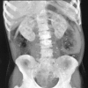
Hemangiomas and vascular malformations of the gastrointestinal tract, rare clinical entities, present as overt or occult bleeding. They can be distributed throughout the intestinal digestive system, or present as a singular cavernous hemangioma or malformation, which is often located in the rectosigmoid region. Misdiagnosis is common despite characteristic radiographic features such as radiolucent phleboliths on plain film imaging and a purplish nodule on endoscopy. Adjunctive imaging such as computed tomography and magnetic resonance imaging are suggested as there is potential for local invasion. Endorectal ultrasound with Doppler has also been found to be useful in some instances. Surgical resection is the mainstay of treatment, with an emphasis on sphincter preservation. Nonsurgical endoscopic treatment with banding and sclerotherapy has been reported with success, especially in instances where an extensive resection is not feasible.
Keywords: Hemangioma; blue rubber bleb; cavernous hemangioma; vascular malformation.
Figures


References
-
- Phillips B. Surgical cases. London Med Gaz. 1839;23:514–517.
-
- Gentry R W, Dockerty M B, Glagett O T. Vascular malformations and vascular tumors of the gastrointestinal tract. Surg Gynecol Obstet. 1949;88(4):281–323. - PubMed
-
- Varma J D, Hill M C, Harvey L AC. Hemangioma of the small intestine manifesting as gastrointestinal bleeding. Radiographics. 1998;18(4):1029–1033. - PubMed
-
- Lyon D T, Mantia A G. Large-bowel hemangiomas. Dis Colon Rectum. 1984;27(6):404–414. - PubMed
-
- Allred H W, Jr, Spencer R J. Hemangiomas of the colon, rectum, and anus. Mayo Clin Proc. 1974;49(10):739–741. - PubMed
LinkOut - more resources
Full Text Sources
Other Literature Sources

