Review
. 2012 Sep-Oct;1(5):693-713.
doi: 10.1002/wdev.52.

Genetic controls and cellular behaviors in branching morphogenesis of the renal collecting system

Affiliations
Review

Genetic controls and cellular behaviors in branching morphogenesis of the renal collecting system

Frank Costantini. Wiley Interdiscip Rev Dev Biol. 2012 Sep-Oct.

Abstract

The mammalian kidney, which at maturity contains thousands of nephrons joined to a highly branched collecting duct (CD) system, is an important model system for studying the development of a complex organ. Furthermore, congenital anomalies of the kidney and urinary tract, often resulting from defects in ureteric bud branching morphogenesis, are relatively common human birth defects. Kidney development is initiated by interactions between the nephric duct and the metanephric mesenchyme, leading to the outgrowth and repeated branching of the ureteric bud epithelium, which gives rise to the entire renal CD system. Meanwhile, signals from the ureteric bud induce the mesenchyme cells to form the nephron epithelia. This review focuses on development of the CD system, with emphasis on the mouse as an experimental system. The major topics covered include the origin and development of the nephric duct, formation of the ureteric bud, branching morphogenesis of the ureteric bud, and elongation of the CDs. The signals, receptors, transcription factors, and other regulatory molecules implicated in these processes are discussed. In addition, our current knowledge of cellular behaviors that are controlled by these genes and underlie development of the collecting system is reviewed.


Figures

Figure 1
Figure 1. Gross organization of the kidney, and development of the nephron
A, Schematic diagram of the gross organization of the newborn mouse kidney, showing a single nephron and associated collecting duct (black dotted box – see B for an enlarged view). The collecting ducts are the mature derivatives of the ureteric bud (UB), whose immature tips are located in the outer nephrogenic zone and continue to branch at this stage (blue dotted box). B, Enlarged diagram of components of the nephron. GL, glomerulus; BC, Bowman’s capsule; PT, proximal tubule; LH, loop of Henle; DT, distal tubule; CT, connecting tubule; CD, collecting duct. C–E, Stages of nephrogenesis and their relationship to the ureteric bud tips. C, Metanephric mesenchyme cells first condense around the UB tips to form the cap mesenchyme(CM). These CM cells are the stem cells that self renew and also give rise to nephron epithelia. D, the CM cells first form aggregates (PA, pretubular aggregates) which convert into epithelial renal vesicles (RV). E, the RV fuses with the UB tip, and folds to form the comma-shaped body (CB); it continues to develop into an S-shaped body (SB), different regions of which begin to differentiate into segments of the mature nephron (BC, PT, DT, and CT). EC, endothelial cells entering the glomerular cleft to form the glomerular tuft of capillaries.
Figure 2
Figure 2. Formation of the nephric duct and ureteric bud
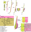
A, Nephric duct (ND), pronephric tubules (PT) and nephrogenic cord (NC) at ~E8.5–E9.0. Enlargements at left: rostral ND has formed an epithelial tube, while caudal ND remains a cord of cells, with filopodia on cells near the caudal tip. B, By E9.5, ND has contacted the cloaca (CL; the precursor of the bladder), and mesonephric tubules (MT) have formed rostrally. C, at E10.0 – E10.5 the metanephric mesenchyme (MM) has formed from the nephrogenic cord, and the adjacent region of the ND has swollen and formed a pseudostratified epithelium, shown in the enlargement. D, At E11.0 the ureteric bud (UB) has emerged and extended into the MM. E, Genes involved in the development of the ND. F, Proteins that promote normal UB formation and outgrowth (lower box) or suppress ectopic budding from the more rostral ND (upper box). Protein symbols in green promote budding while those in red oppose budding (RET continues to be regulated during UB formation, through the same pathways shown in panel E). In the rostral ND, GDNF expression is repressed (indicated by gray lettering) by FOXC1/2 and ROBO2, and thus fails to stimulate RET signaling. Arrows indicate a positive effect (but not necessarily a direct interaction), serial arrows indicate an indirect effect, and dotted arrows an effect that is blocked by an inhibitor (e.g., SPRY1 or GREM1).
Figure 3
Figure 3. Signaling pathways and genes that control UB branching morphogenesis downstream of RET and other RTKs
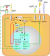
GDNF from the MM binds to the co-receptor GFRA1 on UB cells, and the GDNF/ G FRA1 complex binds to RET, leading to RET dimerization (not depicted) and auto-phosphorylation on several RET tyrosine residues (pY). PLCγ binds to RET pY1015, while adapter proteins (e.g., SHC, GRB2) bind to other pY residues, leading to activation of the PI3K and ERK MAP kinase signaling pathways (as well as others not shown) , . RET signaling upregulates many genes (blue box) in UB tip cells . Genes in green type promote UB growth and branching, those in red oppose it, and those in gray lack a known role. Etv4 and Etv5 are upregulated by RET via PI3K, and are in turn required for normal expression of Cxcr4, Met, Mmp14 and Myb; Etv4 and Etv5 are not required (or are less important) for expression of Dusp6, Spred2, Spry1, Ret and Wnt11. Sox8 and Sox9, which may act downstream of RET, are required for expression of Etv4 and Etv5, as well as Cxcr4, Dusp6, Met, and Spry1 . In positive feed-forward loops (green arrows), RET up-regulates its own expression and that of Wnt11, which up-regulates Gdnf expression in the MM (via an unknown mechanism); in negative feedback loops (red arrows), RET up-regulates expression of Dusp6, Spred2 and Spry1, all negative regulators of RTK signaling). Dashed lines indicate indirect effects or interactions. In addition to RET, several other RTKs including FGFR2, MET (HGF receptor) and EGFR also promote UB growth and branching, and signal through the same major pathways as RET.
Figure 4
Figure 4. Chimera analysis of the effects of RET signaling defects on ND and UB cell behavior in organ cultures
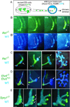
A, ES cells with mutations in Ret, Etv4 and Etv5, or Spry1 (all carrying a Hoxb7/GFP transgene) were injected into blastocysts of a strain expressing CFP in the ND/UB lineage. Thus, in the chimeric embryos, mutant ND or UB cells are GFP+ and WT cells are CFP+. Panels from left to right show successive stages of ND development (B, B’), or UB outgrowth and branching (C–E). B and B’, development in culture of a Ret−/−↔WT chimera from ~E10.0. B shows CFP and GFP, revealing interspersion of WT and Ret−/− cells in the ND at 0 hr, but enrichment of WT cells at the primary UB tip domain (arrow) and the common nephric duct (CND, *) by 24 hr. B’ shows only the CFP channel of the same specimen, revealing that WT cells are rearranged to form the UB tip domain (brackets) and the CND (*). C, in Ret−/−↔WT chimeras at later stages of UB formation, outgrowth and branching, Ret−/− cells contribute to the UB trunks, but not to the tips. D, Etv4−/−, Etv5+/− cells behave similarly to Ret−/− cells in the UB, although they contribute to the CND (*). E, Spry1−/− cells behave oppositely to Ret−/− cells, contributing preferentially to the UB tips (formation of multiple UBs is a common property of Spry1−/− embryos ). Modified from refs., .
Figure 5
Figure 5. Cell migration during mouse ureteric bud and zebrafish pronephric duct development
A–C, model of ND and UB cell movements during formation of the primary UB tip domain, based on analysis of chimeric embryos (Figure 4). Cells in the ND with higher RET signaling activity (blue) preferentially move (A, yellow arrows) to the dorsal ND, adjacent to the MM (pink oval), then form the first UB tip as it emerges (B, C). Cells with lower RET activity (green) do not participate in tip formation, but later contribute to the UB trunk (C, red arrows). D–F, collective cell migration during zebrafish pronephric duct development . D, schematic diagram of zebrafish larva showing pronephric duct (green, PND) and glomerulus (blue). E, schematic diagram of a segment of pronephric duct, indicating movement of ductal cells towards the anterior (pink arrows). Pronephric duct cells migrate collectively, retaining their relative positions, rather than sorting into different domains like mouse nephric duct cells . F, enlargement of dotted box in E shows cryptic lamellipodia, cellular extensions along the basement membrane (BM) thought to be involved in cell movement .
Figure 6
Figure 6. Ureteric bud branching in organ culture and in vivo
A–F, UB branching during culture of a Hoxb7/eGFP transgenic kidney. A, E11.5 kidney at beginning of culture. GFP labels the ND and UB, which has branched once at this stage (“T-stage”), while the surrounding MM is invisible. B, at 10 hours of culture, the two UB tips have trifurcated to generate six tips (yellow stars). C–F, branching patterns at the indicated times. Orange stars in D and E indicate a lateral branch. G, Optical section of a UB tip at E11.5 (similar plane of section to the green dotted line in A). The kidney carries Hoxb7/myrVenus, a transgene encoding a membrane-bound fluorescent protein that reveals the pseudostratified epithelium . H, Optical section of a branching UB tip at E14.5 (a similar stage of branching to that indicated by the green box in E). Note the continuous lumen and the simple, cuboidal epithelium. G and H were reproduced from reference . I–N, Mouse kidneys at the indicated stages of in vivo development, stained with anti-pan-cytokeratin antibody to visualize the ureteric bud tree, and imaged by optical projection tomography and 3D rendering (reproduced from Short et al., 2010 by permission of the authors). Each panel shows the kidney from two perspectives rotated 90 degrees. Note the three dimensional nature of the kidney in vivo, in contrast to the flattened UB tree that develops in organ culture. The inset (M’) shows examples of orthogonal bifurcation. O–Q, Serial sections showing connection of E15.5 UB tip (labeled with anti-Calbindin antibody, green) to a nascent nephron. The basement membrane (labeled with anti-collagen IV, in red) is discontinuous where the nephron joins the UB (yellow stars). Images in O–Q provided by Kylie Georgas, from ref. .
Figure 7
Figure 7. Collecting duct elongation gives rise to the renal medulla and papilla
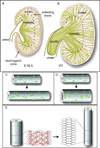
After the phase of rapid UB branching (~E11.5 – E14.5), internal elongation of the CDs generates the medulla (A), first visible at ~E15.5, and the papilla (B), which elongates extensively after birth. C, Oriented cell division along the ductal axis normally contributes to the elongation of CDs. D, In mutants where oriented cell division is randomized, the ducts increase in diameter rather than lengthen, preventing normal development of the medulla and papilla. E, another cellular mechanism of CD elongation appears to be cellular intercalation. See text for references.

References

    1. Cebrian C, Borodo K, Charles N, Herzlinger DA. Morphometric index of the developing murine kidney. Dev Dyn. 2004;231:601–608. - PubMed
    1. Hoy WE, Bertram JF, Denton RD, Zimanyi M, Samuel T, Hughson MD. Nephron number, glomerular volume, renal disease and hypertension. Curr Opin Nephrol Hypertens. 2008;17:258–265. - PubMed
    1. Dressler GR. Advances in early kidney specification, development and patterning. Development. 2009;136:3863–3874. - PMC - PubMed
    1. Levinson RS, Batourina E, Choi C, Vorontchikhina M, Kitajewski J, Mendelsohn CL. Foxd1-dependent signals control cellularity in the renal capsule, a structure required for normal renal development. Development. 2005;132:529–539. - PubMed
    1. Cullen-McEwen LA, Caruana G, Bertram JF. The where, what and why of the developing renal stroma. Nephron Exp Nephrol. 2005;99:e1–e8. - PubMed

Publication types

LinkOut - more resources