

doi: 10.3978/j.issn.2078-6891.2012.021.
Gastric cancer: Classification, histology and application of molecular pathology
- PMID: 22943016
- PMCID: PMC3418539
- DOI: 10.3978/j.issn.2078-6891.2012.021
Item in Clipboard
Gastric cancer: Classification, histology and application of molecular pathology
J Gastrointest Oncol.
2012 Sep.
Abstract
Gastric cancer remains one of the deadly diseases with poor prognosis. New classification of gastric cancers based on histologic features, genotypes and molecular phenotypes helps better understand the characteristics of each subtype, and improve early diagnosis, prevention and treatment. The objective of this article is to review the new classification of gastric cancers and the up-to-date guidance in the application of molecular testing.
Keywords: CDH1; DPD; Gastric carcinoma; HER2; classification; histology; molecular pathology.
Figures
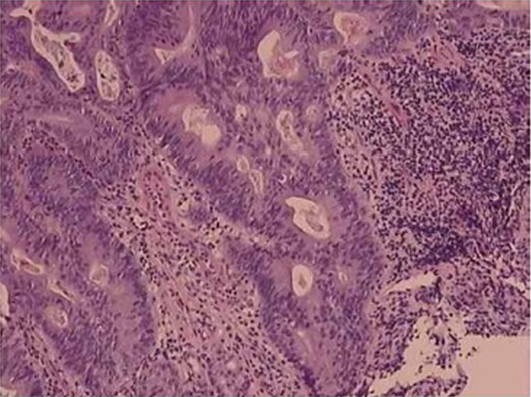
Tubular adenocarcinoma. Irregular-shaped and fused neoplastic glands with intraluminal mucus and debris
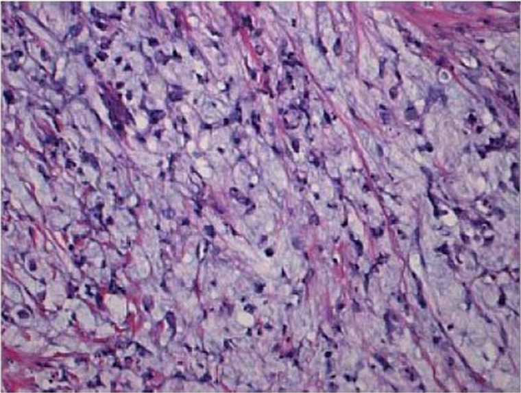
Mucinous adenocarcinoma. Clusters and scattered tumor cells floating in the abundant extracellular mucin pools
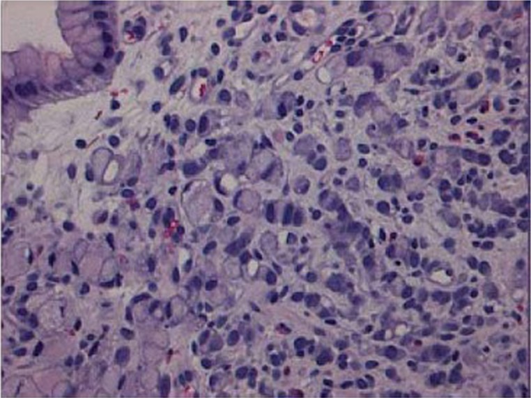
Signet ring cell carcinoma. Signet ring carcinoma cells are predominantly at the superficial lamina propria
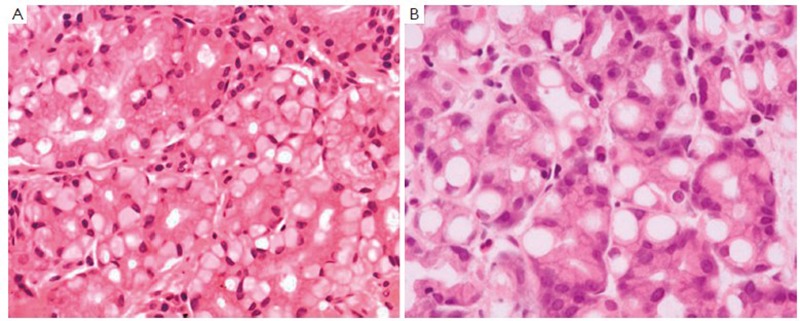
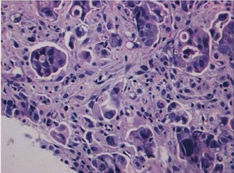
Pseudo-signet ring cells. The cytoplasm of pseudo-signet ring cells are vacuolated (A) and pale (B) (photos are courtesy of Dr. Caroline Hughes)
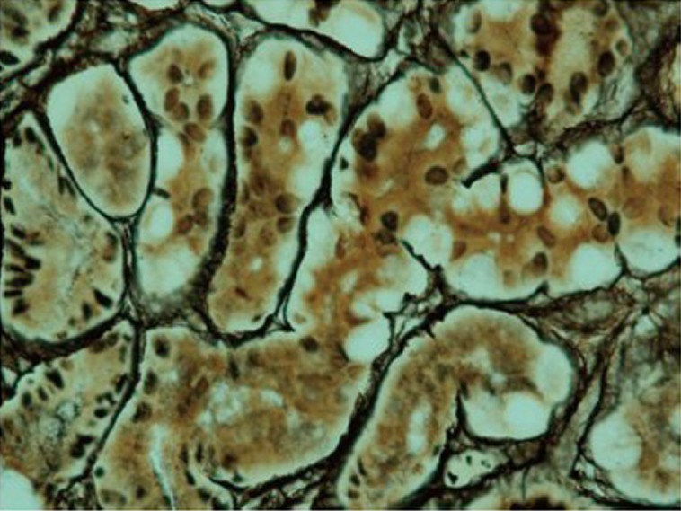
Pseudo-signet ring cells are confined within basement membrane and maintain intact acinar structure with reticulin stain (photo is courtesy of Dr. Caroline Hughes)
Micropapilary adenocarcinoma. Small papillary clusters of tumor cells devoid of fibrovascular core and surrounded by empty spaces

A.In situ signet ring carcinoma cells confined within basement membrane; B. Pagetoid spread of signet ring cells (arrow heads) below the preserved surface epithelium; C. Focus of intramucosal signet ring cell carcinoma (arrows) in the lamina propria (all three photos are courtesy of Dr. Rebecca Fitzgerald)
References
-
- Parkin DM. International variation. Oncogene 2004;23:6329-40 - PubMed
-
- Ferlay J, Shin HR, Bray F, et al. Estimates of worldwide burden of cancer in 2008: GLOBOCAN 2008. Int J Cancer 2010;127:2893-917 - PubMed
-
- Devesa SS, Blot WJ, Fraumeni JF., Jr Changing patterns in the incidence of esophageal and gastric carcinoma in the United States. Cancer 1998;83:2049-53 - PubMed
-
- Blot WJ, Devesa SS, Kneller RW, et al. Rising incidence of adenocarcinoma of the esophagus and gastric cardia. JAMA 1991;265:1287-9 - PubMed
-
- Jemal A, Bray F, Center MM, et al. Global cancer statistics. CA Cancer J Clin 2011;61:69-90 - PubMed
LinkOut - more resources
Full Text Sources
Research Materials
Miscellaneous