

Characterization of a distinct lethal arteriopathy syndrome in twenty-two infants associated with an identical, novel mutation in FBLN4 gene, confirms fibulin-4 as a critical determinant of human vascular elastogenesis
- PMID: 22943132
- PMCID: PMC3598868
- DOI: 10.1186/1750-1172-7-61
Characterization of a distinct lethal arteriopathy syndrome in twenty-two infants associated with an identical, novel mutation in FBLN4 gene, confirms fibulin-4 as a critical determinant of human vascular elastogenesis
Abstract
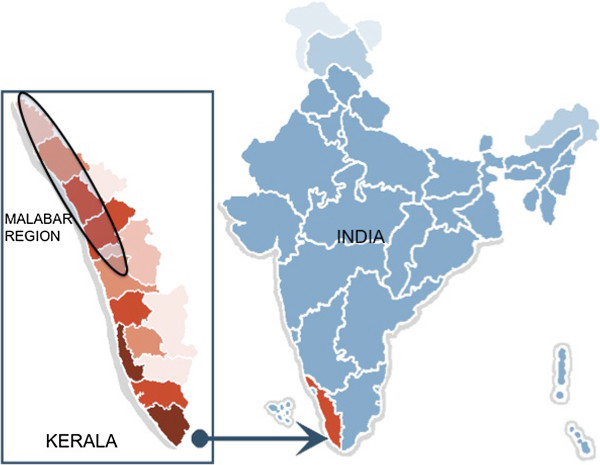
Background: Vascular elasticity is crucial for maintaining hemodynamics. Molecular mechanisms involved in human elastogenesis are incompletely understood. We describe a syndrome of lethal arteriopathy associated with a novel, identical mutation in the fibulin 4 gene (FBLN4) in a unique cohort of infants from South India.
Methods: Clinical characteristics, cardiovascular findings, outcomes and molecular genetics of twenty-two infants from a distinct population subgroup, presenting with characteristic arterial dilatation and tortuosity during the period August 2004 to June 2011 were studied.
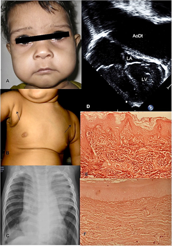
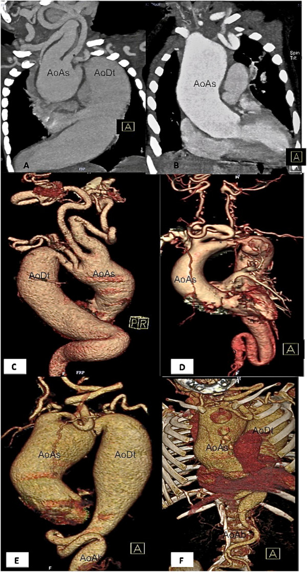
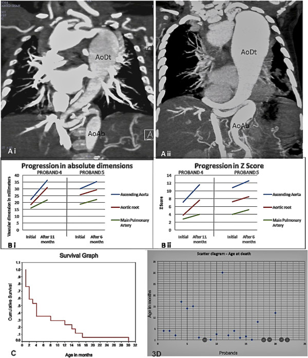
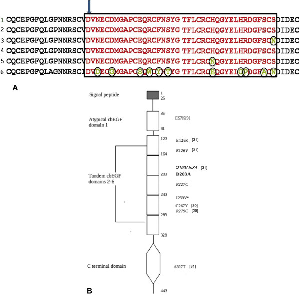
Results: Patients (11 males, 11 females) presented at median age of 1.5 months, belonging to unrelated families from identical ethno-geographical background; eight had a history of consanguinity. Cardiovascular features included aneurysmal dilatation, elongation, tortuosity and narrowing of the aorta, pulmonary artery and their branches. The phenotype included a variable combination of cutis laxa (52%), long philtrum-thin vermillion (90%), micrognathia (43%), hypertelorism (57%), prominent eyes (43%), sagging cheeks (43%), long slender digits (48%), and visible arterial pulsations (38%). Genetic studies revealed an identical c.608A > C (p. Asp203Ala) mutation in exon 7 of the FBLN4 gene in all 22 patients, homozygous in 21, and compound heterozygous in one patient with a p. Arg227Cys mutation in the same conserved cbEGF sequence. Homozygosity was lethal (17/21 died, median age 4 months). Isthmic hypoplasia (n = 9) correlated with early death (≤4 months).
Conclusions: A lethal, genetic disorder characterized by severe deformation of elastic arteries, was linked to novel mutations in the FBLN4 gene. While describing a hitherto unreported syndrome in this population subgroup, this study emphasizes the critical role of fibulin-4 in human elastogenesis.
Figures
References
-
- Coucke PJ, Willaert A, Wessels MW, Callewaert B, Zoppi N, De Backer J, Fox JE, Mancini GM, Kambouris M, Gardella R, Facchetti F, Willems PJ, Forsyth R, Dietz HC, Barlati S, Colombi M, Loeys B, De Paepe A. Mutations in the facilitative glucose transporter GLUT10 alter angiogenesis and cause arterial tortuosity syndrome. Nat Genet. 2006;38(4):452–457. doi: 10.1038/ng1764. - DOI - PubMed
-
- Loeys BL, Chen J, Neptune ER, Judge DP, Podowski M, Holm T, Meyers J, Leitch CC, Katsanis N, Sharifi N. Xu Fl, Myers LA, Spevak PJ, Cameron DE, De Backer J, Hellemans J, Chen Y, Davis EC, Webb CL, Kress W, Coucke P, Rifkin DB, De Paepe AM, Dietz HC. A syndrome of altered cardiovascular, craniofacial, neurocognitive and skeletal development caused by mutations in TGFBR1 or TGFBR2. Nat Genet. 2005;37(3):275–281. doi: 10.1038/ng1511. - DOI - PubMed
-
- Al Fadley F, Al Manea W, Nykanen DG, Al Fadley A, Bulbul Z, Al Halees Z. Severe tortuosity and stenosis of the systemic, pulmonary and coronary vessels in 12 patients with similar phenotypic features: a new syndrome? Cardiol Young. 2000;10(6):582–589. - PubMed
-
- Wessels MW, Catsman-Berrevoets CE, Mancini GM, Breuning MH, Hoogeboom JJ, Stroink H, Frohn-Mulder I, Coucke PJ, Paepe AD, Niermeijer MF, Willems PJ. Three new families with arterial tortuosity syndrome. Am J Med Genet A. 2004;131(2):134–143. - PubMed
Publication types
MeSH terms
Substances
LinkOut - more resources
Full Text Sources
Medical
Molecular Biology Databases
