. 2012 Dec 1;448(2):189-200.
doi: 10.1042/BJ20120339.

2-APB-potentiated channels amplify CatSper-induced Ca(2+) signals in human sperm

Affiliations

2-APB-potentiated channels amplify CatSper-induced Ca(2+) signals in human sperm

Linda Lefièvre et al. Biochem J. .

Abstract

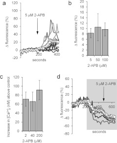
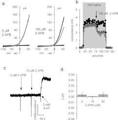
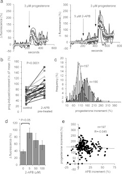
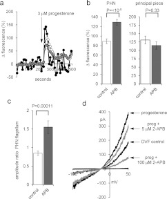
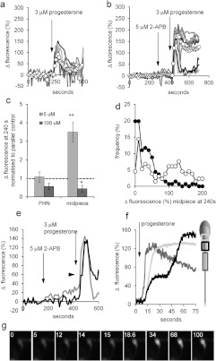
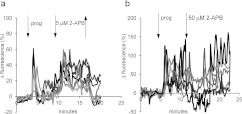
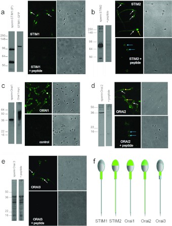
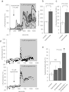
Ca2+i signalling is pivotal to sperm function. Progesterone, the best-characterized agonist of human sperm Ca2+i signalling, stimulates a biphasic [Ca2+]i rise, comprising a transient and subsequent sustained phase. In accordance with recent reports that progesterone directly activates CatSper, the [Ca2+]i transient was detectable in the anterior flagellum (where CatSper is expressed) 1-2 s before responses in the head and neck. Pre-treatment with 5 μM 2-APB (2-aminoethoxydiphenyl borate), which enhances activity of store-operated channel proteins (Orai) by facilitating interaction with their activator [STIM (stromal interaction molecule)] 'amplified' progesterone-induced [Ca2+]i transients at the sperm neck/midpiece without modifying kinetics. The flagellar [Ca2+]i response was unchanged. 2-APB (5 μM) also enhanced the sustained response in the midpiece, possibly reflecting mitochondrial Ca2+ accumulation downstream of the potentiated [Ca2+]i transient. Pre-treatment with 50-100 μM 2-APB failed to potentiate the transient and suppressed sustained [Ca2+]i elevation. When applied during the [Ca2+]i plateau, 50-100 μM 2-APB caused a transient fall in [Ca2+]i, which then recovered despite the continued presence of 2-APB. Loperamide (a chemically different store-operated channel agonist) enhanced the progesterone-induced [Ca2+]i signal and potentiated progesterone-induced hyperactivated motility. Neither 2-APB nor loperamide raised pHi (which would activate CatSper) and both compounds inhibited CatSper currents. STIM and Orai were detected and localized primarily to the neck/midpiece and acrosome where Ca2+ stores are present and the effects of 2-APB are focussed, but store-operated currents could not be detected in human sperm. We propose that 2-APB-sensitive channels amplify [Ca2+]i elevation induced by progesterone (and other CatSper agonists), amplifying, propagating and providing spatio-temporal complexity in [Ca2+]i signals of human sperm.


Figures

Figure 1
Figure 1. 2-APB elevates resting [Ca2+]i
(a) 2-APB (5 μM; arrow) causes a sustained increase in the PHN [Ca2+]i of a subset of cells. The traces show ten individual cell responses and ΔFmean (○-○) for all 87 cells in the experiment. (b and c) 2-APB-induced [Ca2+]i elevation is dose-independent. (b) Increase in ΔFmean 3 min after application of 2-APB. Results are means±S.E.M. for sets of four experiments in each of which aliquots from the sample were tested with each of the three concentrations of 2-APB. (c) Dose-dependence of 2-APB-induced [Ca2+]i increment in fura-2-loaded cell populations (means±S.E.M. for 6–17 experiments). (d) 2-APB-induced rise in [Ca2+]i is reversed in low-Ca2+ saline. Cells were superfused with EGTA-buffered saline (shown by shading) then exposed to 5 μM 2-APB (arrow). 2-APB-induced [Ca2+]i increase was abolished and in many cells 2-APB induced a further fall in [Ca2+]i. Traces show six individual cell responses and ΔFmean (○-○) for all 85 cells in the experiment.
Figure 2
Figure 2. 2-APB does not enhance CatSper currents
(a) Monovalent CatSper currents recorded before (black trace) and after (grey trace) application of 5 μM (left-hand panel) and 100 μM (right-hand panel) 2-APB. Horizontal (near zero) traces show currents in divalent cation-containing saline. (b) Time course of CatSper current block by 100 μM 2-APB. Cell conductance was calculated from the slope between +50 and +60 mV. Grey shading shows superfusion with DVF saline, arrow shows application of 2-APB. (c) 2-APB does not raise pHi. 2-APB at 5 and 15 μM was added at the first and second arrows respectively. 4-Aminopyridine (2 mM; 4-AP; positive control) caused an immediate rise in pHi. (d) Mean pHi change (±S.E.M.) in response to 5 μM (n=6), 15 μM (n=3) and 50 μM (n=3) 2-APB.
Figure 3
Figure 3. 2-APB modulates the progesterone-induced [Ca2+]i transient
(a) Elevation of [Ca2+]i at the PHN in response to stimulation with 3 μM progesterone (arrow) under control conditions (left-hand panel) and after 200 s exposure to 5 μM 2-APB (right-hand panel). Both experiments used cells from the same preparation. Traces show 6–8 representative single-cell responses and ΔFmean (○-○) for all 107 (left-hand panel) and 125 (right-hand panel) cells in the experiment. (b) Summary of results from 20 pairs of control and 5 μM 2-APB-pre-treated experiments. Each point shows the mean amplitude of the progesterone-induced transient (increment in ΔFmean) for all of the cells in a single experiment (50–200 cells). Joined pairs of points show 5 μM 2-APB pre-treatment (right-hand point) and corresponding control (left-hand point) using cells from the same ejaculate – such as the pair shown in (a). Data from 20 pairs of experiments are shown and the overall mean for all 20 is shown by Δ-Δ. (c) Effect of 5 μM 2-APB on amplitude distribution of single-cell progesterone-induced transients. Grey bars show the control, black bars show the parallel 5 μM 2-APB-pre-treated experiment. (d) Dose-dependence of potentiation by 2-APB of the progesterone-induced [Ca2+]i transient. Each bar shows the mean amplitude±S.E.M. for four sets of experiments (50–200 cells each). In each set, four experiments were carried out with samples from the same ejaculate, using 0, 5, 50 or 100 μM 2-APB applied 200 s before progesterone. Only 5 μM 2-APB significantly enhanced the [Ca2+]i transient. (e) Amplitude of 2-APB-induced resting [Ca2+]i elevation (APB increment; x-axis) is not correlated with the amplitude of subsequent progesterone-induced [Ca2+]i transient (progesterone increment; y-axis). Results are from 197 cells in one experiment.
Figure 4
Figure 4. 2-APB does not enhance the flagellar Ca2+ signal or potentiate activation of CatSper by progesterone
(a) [Ca2+]i (OGB) signal from the PHN (white circles), midpiece (grey circles) and flagellum (black circles) in response to application of 3 μM progesterone (arrow). Each trace shows the mean response from the same nine cells. (b) Amplitude of progesterone-induced [Ca2+]i transient at the PHN (left-hand panel) and midpiece (right-hand panel) under control conditions (white bars; n=43 cells) and after pre-treatment with 5 μM 2-APB (grey bars; n=57 cells). (c) Ratio of [Ca2+]i transient amplitudes simultaneously recorded from the PHN and flagellum under control conditions (white bar; n=43) and after pre-treatment with 5 μM 2-APB (grey bar; n=57). (d) Monovalent currents (DVF control) were enhanced by 500 nM progesterone (upper black trace). Subsequent application of 5 μM 2-APB (upper grey trace) and 100 mM 2-APB (lower grey trace) reduced the amplitude of outward and inward currents.
Figure 5
Figure 5. 2-APB (5 μM) enhances sustained elevation of [Ca2+]i in the midpiece
(a and b) Progesterone-induced responses at the midpiece under control conditions (a) and after application of 5 μM 2-APB (first arrow; b). Each plot shows six to nine representative single-cell traces and ΔFmean (○-○) for all 25 (a) and 31 (b) cells in the experiment. (c) Dose-dependence of the effect of 2-APB pre-treatment on the sustained [Ca2+]i signal. The amplitude of the sustained response (240 s after progesterone addition) in PHN (left-hand bars) and midpiece (right-hand bars) after exposure to 5 μM 2-APB (light grey bars) and 100 μM 2-APB (dark grey bars) was normalized to the amplitude of the parallel control (shown by a broken line). Each bar shows the means±S.E.M. of 11 (5 μM 2-APB) and six (100 μM 2-APB) experiments. *P<0.05; **P<0.005 compared with control. (d) Amplitude distribution of single-cell sustained [Ca2+]i increases (240 s after progesterone addition). Open circles (○-○) show responses of 2-APB pre-treated cells (136 cells from five experiments), closed circles (●-●) show responses from 135 cells in the five parallel control experiments. (e) Progesterone-stimulated [Ca2+]i elevation in the PHN (grey trace) and midpiece (black trace) of a 5 μM 2-APB pre-treated cell. 2-APB was added at the first arrow, 3 μM progesterone at the second arrow. An inflection in the rising phase of the midpiece trace occurs ~20 s after application of progesterone (arrowhead). (f) Progesterone-stimulated [Ca2+]i elevation in the anterior flagellum (dark grey), PHN (light grey) and midpiece (black) of a 5 μM 2-APB pre-treated cell imaged at 10 Hz. Progesterone was applied at 7 s (arrow). The anterior flagellar response precedes responses in the other two compartments and the rising phase of the midpiece response shows an inflexion ~25 s after onset. (g) Image series of the same cell as (f), showing the delayed [Ca2+]i rise in the midpiece. Numbers show time in seconds. Progesterone was applied at 7 s.
Figure 6
Figure 6. 2-APB modifies the sustained [Ca2+]i elevation
(a) 2-APB (5 μM) was applied to cells already stimulated with 3 μM progesterone (prog). 2-APB caused a tonic increase in [Ca2+]i that reversed upon washout (↑) of the drug. Traces show PHN responses from seven individual representative cells. (b) 2-APB (50 μM) was applied to cells already stimulated with 3 μM progesterone. Upon application of the drug, [Ca2+]i fell and oscillations were suppressed, but [Ca2+]i then recovered despite the continued presence of the drug. Traces show PHN responses from six individual representative cells.
Figure 7
Figure 7. Expression of Orai and STIM in human sperm
(a) STIM1. Left-hand panels: Western blot for STIM1 (ProSci 4119); lane 1: human sperm proteins purified by immunoprecipitation with an anti-STIM1 antibody. A band is seen at ~95 kDa and also at 55–60 kDa due to the presence of anti-STIM1 antibody from the immunoprecipitation procedure. Lane 2 is protein from STIM1–GFP-transfected HEK-293 cells. STIM1 appears at ~110 kDa due to the presence of the 25 kDa GFP tag. Separation of images in this and other gels indicates that lanes were not originally directly adjacent. Right-hand panels: immunofluorescent staining with anti-STIM1 antibody (ProSci). Upper panels show STIM1 staining and the corresponding phase image. Fluorescence occurs over the midpiece with a bright spot at the sperm neck (arrows). Lower panels show cells incubated with antibody pre-adsorbed with the antigenic peptide, which abolished staining. (b) STIM2. Left-hand panels: Western blot for STIM2 (ProSci antibody 4123); lane 1: human sperm proteins. An intense doublet is present at 85–90 kDa. Lane 2: as lane 1, but antibody was pre-adsorbed with the antigenic peptide. Right-hand panels: immunofluorescent staining with anti-STIM2 antibody. The upper panels show STIM2 staining and corresponding phase image. Staining occurs over the flagellum, being heaviest at the midpiece (white arrows). In a minority of cells (<10%), we observed staining over the acrosome (yellow arrow). The lower panels show cells incubated with antibody pre-adsorbed with the antigenic peptide, which abolished flagellar and acrosomal staining but resulted in fluorescence just behind the equatorial segment (blue arrows). (c) Orai 1. Left-hand panels: Orai 1 immunoblot (Sigma antibody O8264); lane 1: human sperm proteins. Lane 2: proteins extracted from Myc-tagged Orai 1-transfected HEK-293 cells. Deduced molecular mass of non-glycosylated Orai 1 is ~35 kDa. Right-hand panel: immunofluorescent staining with anti-Orai 1 antibody. Upper panels show Orai 1 staining (Sigma antibody O8264) and corresponding phase image. Staining occurs primarily over the acrosome and midpiece and weakly on the principal piece. Lower panels show cells stained similarly but omitting the primary antibody. (d) Orai 2. Left-hand panels: Western blot for Orai 2 (ProSci antibody 4111); lane 1: human sperm proteins. Lane 2: as lane 1, but antibody pre-adsorbed with the antigenic peptide. Right-hand panel: immunofluorescent staining with anti-Orai 2 antibody. Upper panels show Orai 2 staining and corresponding phase image. Staining occurs over the midpiece (white arrows) and acrosome (yellow arrows), with weaker staining over the principal piece. Lower panels show cells incubated with antibody pre-adsorbed with the antigenic peptide, which reduces/abolishes staining of the acrosome, midpiece and flagellum, but resulted in fluorescence just behind the equatorial segment (blue arrows). (e) Orai 3. Left-hand panels: Western blot for Orai 3 (ProSci antibody 4215). Lane 1: human sperm proteins. Lane 2: as lane 1, but antibody was pre-adsorbed with the antigenic peptide, which did not block band detection. Right-hand panels: immunofluorescent staining with anti-Orai 3 antibody. Upper panels show Orai 3-staining and corresponding phase image. Staining occurs primarily over the anterior midpiece and sperm neck (arrows). Lower panels show cells incubated with antibody pre-adsorbed with the antigenic peptide, which abolished staining. (f) Diagrammatic representation of ‘typical’ localization (immunofluorescence pattern) for each of the proteins investigated.
Figure 8
Figure 8. Loperamide potentiates the response of human sperm to progesterone
(a) Pre-treatment with 10 μM loperamide (arrow) followed by application of 3 μM progesterone (shading). Traces show nine representative single-cell PHN responses and ΔFmean (○-○) for all 81 cells in the experiment. Loperamide elevates resting [Ca2+]i and subsequent exposure to progesterone induced an initial [Ca2+]i transient followed by large [Ca2+]i oscillations. (b) Duration of the progesterone-induced [Ca2+]i transient in the PHN was increased by loperamide pre-treatment. Bars show means±S.E.M. for 11 paired experiments. (c) Progesterone-induced sustained [Ca2+]i increase (ΔFmean at 240 s after progesterone) was enhanced by loperamide pre-treatment. Bars show means±S.E.M. for nine paired experiments. (d) Mean normalized fluorescence (ΔFmean) in the PHN (○-○) and in the midpiece (●-●) under control conditions (upper panel; mean of 19 cells) and after pre-treatment with 10 μM loperamide (lower panel; mean of 33 cells). Potentiation by loperamide of [Ca2+]i transient duration and sustained [Ca2+]i elevation are similar in the two compartments. (e) Loperamide enhances progesterone-induced hyperactivation. Each bar shows the percentage of hyperactivated cells (means±S.E.M.; n=7). Progesterone (3 μM) and loperamide (10 μM), applied individually, failed significantly to increase hyperactivation (not significant; NS). When cells were pre-treated with loperamide (3 min), progesterone significantly increased the proportion of hyperactivated cells over all the other conditions (*P<0.02).

References

    1. Publicover S., Harper C. V., Barratt C. [Ca2+]i signalling in sperm–making the most of what you’ve got. Nat. Cell Biol. 2007;9:235–242. - PubMed
    1. Kirichok Y., Lishko P. V. Rediscovering sperm ion channels with the patch-clamp technique. Mol. Hum. Reprod. 2011;17:478–499. - PMC - PubMed
    1. Lishko P. V., Kirichok Y., Ren D., Navarro B., Chung J. J., Clapham D. E. The control of male fertility by spermatozoan ion channels. Annu. Rev. Physiol. 2012;74:453–475. - PMC - PubMed
    1. Brenker C., Goodwin N., Weyand I., Kashikar N. D., Naruse M., Krahling M., Muller A., Kaupp U. B., Strunker T. The CatSper channel: a polymodal chemosensor in human sperm. EMBO J. 2012;31:1654–1665. - PMC - PubMed
    1. Ren D., Xia J. Calcium signaling through CatSper channels in mammalian fertilization. Physiology (Bethesda) 2010;25:165–175. - PubMed

Publication types

MeSH terms