

The co-expression of cytokeratin and p63 in epithelioid angiosarcoma of the parotid gland: a diagnostic pitfall
- PMID: 22943673
- PMCID: PMC3487964
- DOI: 10.1186/1746-1596-7-118
The co-expression of cytokeratin and p63 in epithelioid angiosarcoma of the parotid gland: a diagnostic pitfall
Abstract
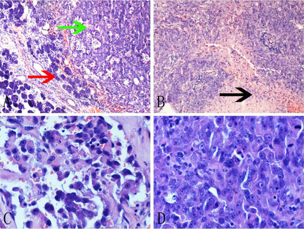
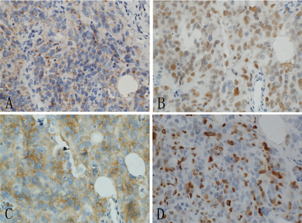
Epithelioid angiosarcoma of the parotid gland is rare, and may pose a great diagnostic challenge. We report a case of primary epithelioid angiosarcoma in a 64-year-old male without history of radiation. The histopathological findings demonstrated a high grade epithelioid neoplasm. Immunostaining showed that the tumor was positive for the pan-cytokeratin, p63, cytokeratin18, Vimentin and vascular markers CD31, and was negative for CD34, cytokeratin5/6, cytokeratin7, cytokeratin20, CD68, CD30, S-100, HMB45, desmin, α-SMA and CD45. The tumor was diagnosed as an epithelioid angiosarcoma. To our knowledge, this is the first reported case of angiosarcoma which showed common positivity for cytokeratin and p63. In addition to cytokeratin, p63 is considered a useful marker for carcinoma. The co-expression of cytokeratin and p63 in epithelioid angiosarcoma represents a diagnostic pitfall. Thus, using a panel of antibodies is essential for distinguishing this tumor from poorly differentiated carcinoma. Virtual slides: The virtual slide(s) for this article can be found here: http://www.diagnosticpathology.diagnomx.eu/vs/6548916707504750.
Figures
References
-
- Fletcher CDM, Unni KK, Mertens F. World Health Organization classification of tumours: pathology and genetics of tumours of soft tissue and bone. Lyon: IARC Press; 2002.
Publication types
MeSH terms
Substances
LinkOut - more resources
Full Text Sources
Research Materials
Miscellaneous
