. 2013;22(2):231-42.
doi: 10.3727/096368912X654939. Epub 2012 Aug 27.

Human-scale whole-organ bioengineering for liver transplantation: a regenerative medicine approach

Affiliations

Human-scale whole-organ bioengineering for liver transplantation: a regenerative medicine approach

Hiroshi Yagi et al. Cell Transplant. 2013.

Abstract

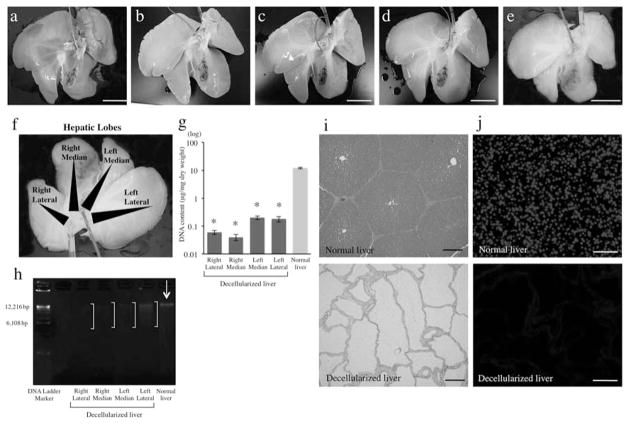
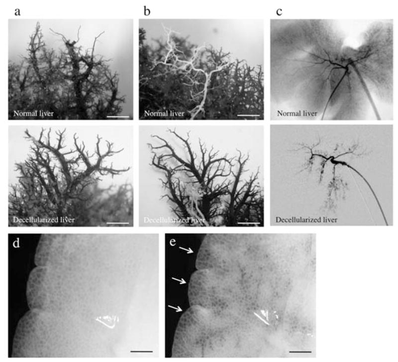
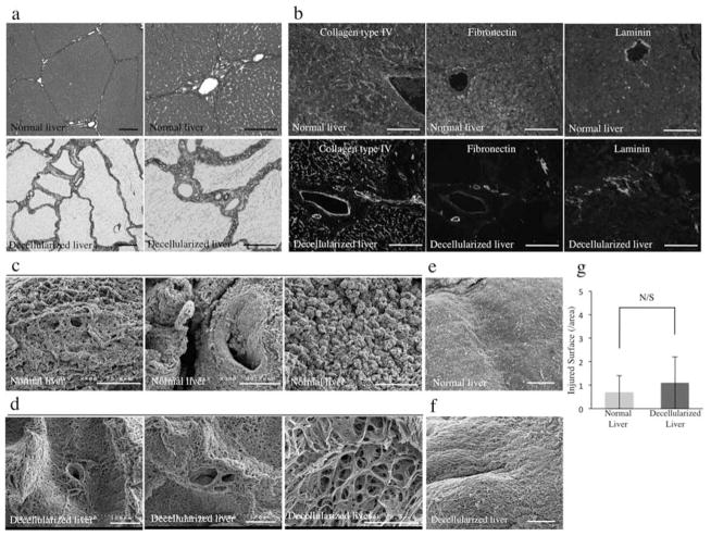
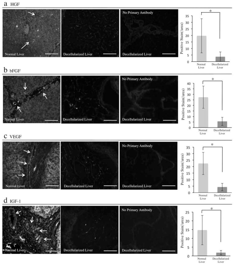
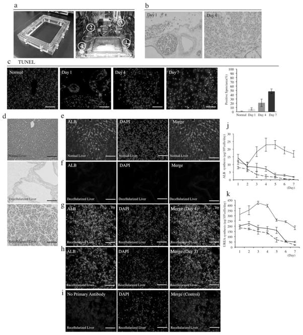
At this time, the only definitive treatment of hepatic failure is liver transplantation. However, transplantation has been limited by the severely limited supply of human donor livers. Alternatively, a regenerative medicine approach has been recently proposed in rodents that describe the production of three-dimensional whole-organ scaffolds for assembly of engineered complete organs. In the present study, we describe the decellularization of porcine livers to generate liver constructs at a scale that can be clinically relevant. Adult ischemic porcine livers were successfully decellularized using a customized perfusion protocol, the decellularization process preserved the ultrastructural extracellular matrix components, functional characteristics of the native microvascular and the bile drainage network of the liver, and growth factors necessary for angiogenesis and liver regeneration. Furthermore, isolated hepatocytes engrafted and reorganized in the porcine decellularized livers using a human-sized organ culture system. These results provide proof-of-principle for the generation of a human-sized, three-dimensional organ scaffold as a potential structure for human liver grafts reconstruction for transplantation to treat liver disease.


Conflict of interest statement

The authors declare no conflicts of interest.

Figures

Figure 1
Figure 1
Whole-organ porcine liver homogeneous decellularization. Representative images of porcine livers during decellularization process at (a) 0 h, (b) 18 h, (c) 48 h, (d) 72 h, and (e) 96 h. (f) DNA was extracted from each different lobe. (g) The DNA content of different lobes of the decellularized liver matrix (n = 4 for each lobe) and (h) agarose gel electrophoresis of extracted DNA comparing to that of normal porcine liver. Histological comparison of normal liver and decellularized liver matrix: (i) hematoxylin and eosin. (j) The presence of intact nuclear material was evaluated by staining the decellularized liver and native liver using 4′,6-diamidino-2-phenylindole (DAPI). *p < 0.01. Scale bars: 5 cm (a–e) and 100 μm (h, i).
Figure 2
Figure 2
Decellularized liver matrix preserves entire vascular system and bile ducts. Representative photographs of decellularized left lobes of porcine liver, with the vascular tree visible. Comparison of normal liver and decellurarized whole-liver bioscaffold of corrosion cast of (a) portal vein (lighter staining) and hepatic artery (darker staining) and (b) hepatic vein and bile duct (white) as well as by (c) radiographic imaging of the bile ducts. (d, e) Representative photographs of visible microvascular tree, after perfusion with heparinized porcine blood through the portal vein. Scale bars: 100 μm.
Figure 3
Figure 3
Morphology. (a) Azan stain of normal porcine livers and decellularized porcine liver scaffolds. (b) Immune-histochemical stain of normal porcine livers (top row) and decellularized porcine liver scaffolds (bottom row) of collagen IV, fibronectin, and laminin. Sections were counterstained with DAPI. (c) SEM images of normal porcine liver and (d) decellularized porcine liver scaffold; representing central vein, portal triad, and extracellular matrix within the parenchyma. (e) Representative photographs of normal and (f) decellularized porcine liver surface (Glisson’s capsule) and (g) the quantification of disruptive areas in the collagenous surface of the livers (n = 4: 10 random images for each sample). Scale bars: 100 and 50 μm (third picture of (c) and (d)). N/S, no significant difference.
Figure 4
Figure 4
Decellularized whole-liver scaffold retained growth factors expression. Immune-histochemical evaluation of growth factors. (a) Hepatocyte growth factor (HGF) stain of native liver and the acellular liver scaffold and its quantification of the positive areas (arrows) per field (n = 4: 10 random images for each sample). (b) Basic fibroblast growth factor (bFGF) stain of native liver and the acellular liver scaffold and its quantification of the positive areas (arrows) per field (n = 4: 10 random images for each sample). (c) Vascular endothelial growth factor (VEGF) stain of native liver and the acellular liver scaffold and its quantification of the positive areas (arrows) per field (n = 4: 10 random images for each sample). (d) Insulin-like growth factor (IGF)-1 stain of native liver and the acellular liver scaffold and its quantification of the positive areas (arrows) per field (n = 4: 10 random images for each sample). Immunostain without primary antibodies with secondary antibodies served as controls in each experiment. Scale bars: 100 μm. *p < 0.05.
Figure 5
Figure 5
Recellularization of a large-scale liver scaffold using porcine primary hepatocytes. (a) Representative photograph of the chamber constructed specifically for a large-scale liver graft and the liver perfusion system (1: closed chamber, 2: peristaltic pump, 3: bubble trap, 4: oxygenator). (b) H&E stain of infused cells in the decellularized liver at day 4 and day 7 (n = 4). (c) Sequential images of terminal deoxynucleotidyl transferase dUTP nick end labeling (TUNEL) stain at different time points and quantitative analysis of TUNEL positive cells (n = 4: 10 random images for each sample). (d) Histological images of normal liver, decellularized liver scaffold, and recellularized liver with primary porcine hepatocytes for H&E stain (n = 4). (e) Albumin (ALB) stain of normal porcine liver, (f) decellularized liver, and recellularized liver with primary porcine hepatocytes after 4 days (g) and 7 days (h) of perfusion. (i) Immunostain without the primary antibody with secondary antibody served as control (n = 4). (j) ALB concentration in the culture medium of collagen-Matrigel sandwich culture (small dashes) or single collagen culture of normal porcine hepatocytes (large dashes) and in the perfusion culture medium of recellularized liver (n = 3). (k) UREA concentration in the culture medium of collagen-Matrigel sandwich culture (small dashes) or single collagen culture of normal porcine hepatocytes (large dashes) and in the perfusion culture medium of recellularized liver (black line) (n = 3). Sections were counterstained with DAPI. Scale bars: 100 μm.

References

    1. Agrawal V, Johnson SA, Reing J, Zhang L, Tottey S, Wang G, Hirschi KK, Braunhut S, Gudas LJ, Badylak SF. Epimorphic regeneration approach to tissue replacement in adult mammals. Proc Natl Acad Sci USA. 2010;107(8):3351–3355. - PMC - PubMed
    1. Avolio AW, Frongillo F, Nicolotti N, Mule A, Vennarecci G, De Simone P, Agnes S. Successful use of extended criteria donor grafts with low to moderate steatosis in patients with model for end-stage liver disease scores below 27. Transplant Proc. 2009;41(1):208–212. - PubMed
    1. Badylak SF, Freytes DO, Gilbert TW. Extracellular matrix as a biological scaffold material: Structure and function. Acta Biomater. 2009;5(1):1–13. - PubMed
    1. Bao J, Shi Y, Sun H, Yin X, Yang R, Li L, Chen X, Bu H. Construction of a Portal implantable functional tissue engineered liver using perfusion-decellularized matrix and hepatocytes in rats. Cell Transplant. 2011;20(5):753–766. - PubMed
    1. Baptista PM, Siddiqui MM, Lozier G, Rodriguez SR, Atala A, Soker S. The use of whole organ decellularization for the generation of a vascularized liver organoid. Hepatology. 2011;53(2):604–617. - PubMed

Publication types