

PRAME expression in head and neck cancer correlates with markers of poor prognosis and might help in selecting candidates for retinoid chemoprevention in pre-malignant lesions
- PMID: 22944049
- PMCID: PMC3607432
- DOI: 10.1016/j.oraloncology.2012.08.005
PRAME expression in head and neck cancer correlates with markers of poor prognosis and might help in selecting candidates for retinoid chemoprevention in pre-malignant lesions
Abstract
Objectives: PRAME (Preferentially Expressed Antigen in Melanoma) is a tumor-associated antigen recognized by immunocytes, and it induces cytotoxic T cell-mediated responses in melanoma. PRAME expression in tumors interferes with retinoic acid receptor (RAR) signaling thus promoting tumor progression. Here, we study PRAME expression in head and neck squamous cell carcinoma (HNSCC) to determine its potential clinical significance.
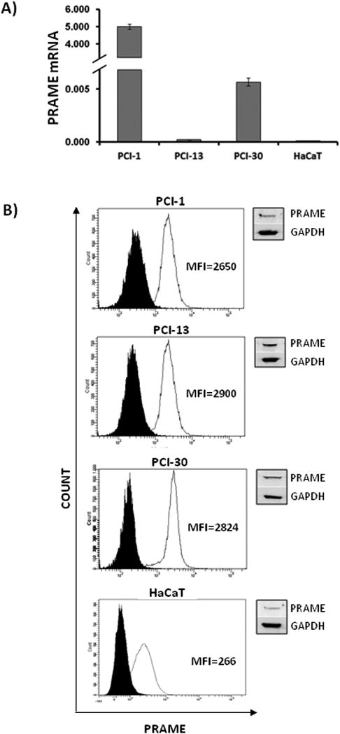
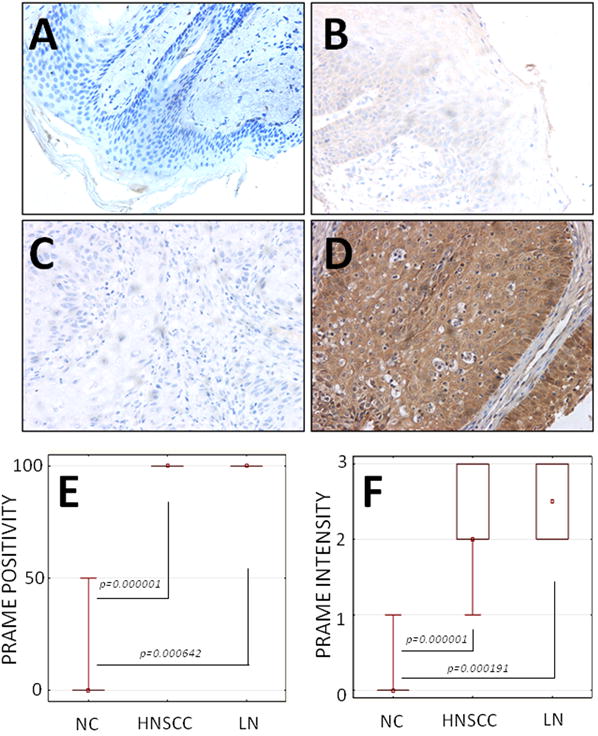
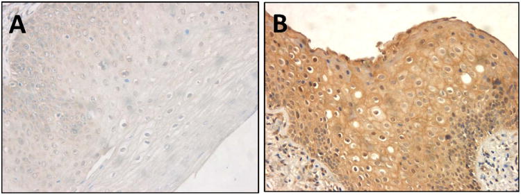
Materials and methods: PRAME expression in HNSCC was evaluated by immunohistochemistry in tissue microarrays of primary tumors (n=53), metastatic lymph nodes (n=8) and normal oral mucosa (n=11). Biopsies of dysplastic oral lesions (n=12) were also examined. PRAME expression levels in tissues were correlated with markers of poor prognosis in HNSCC. PRAME mRNA in HNSCC cell lines and in normal immortalized human keratinocytes (HaCaT cell line) was measured by qRT-PCR, and the protein expression by flow cytometry and western blots.
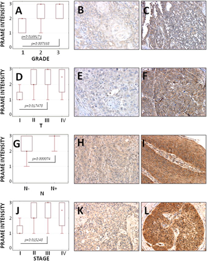
Results: PRAME was expressed in HNSCC cell lines and HNSCC lesions. PRAME expression in dysplastic mucosa was variable. No or only weak expression was found in normal cells or tissues. PRAME expression levels significantly correlated with the tumor grade, size, nodal involvement and the clinical status of HNSCC patients.
Conclusions: Elevated PRAME expression associates with clinicopathologic markers of poor outcome in HNSCC and might identify potential candidates with pre-cancerous lesions for chemoprevention with retinoids.
Copyright © 2012 Elsevier Ltd. All rights reserved.
Figures
References
-
- Forastiere A, Koch W, Trotti A, Sidransky D. Head and neck cancer. N Engl J Med. 2001;345:1890–900. - PubMed
-
- Lorch JH, Goloubeva O, Haddad RI, Cullen K, Sarlis N, Tishler R, et al. Induction chemotherapy with cisplatin and fluorouracil alone or in combination with docetaxel in locally advanced squamous-cell cancer of the head and neck: long-term results of the TAX 324 randomised phase 3 trial. Lancet Oncol. 2011;12:153–9. - PMC - PubMed
-
- Posner MR, Hershock DM, Blajman CR, Mickiewicz E, Winquist E, Gorbounova V, et al. Cisplatin and fluorouracil alone or with docetaxel in head and neck cancer. N Engl J Med. 2007;357:1705–15. - PubMed
-
- Calli C, Calli A, Pinar E, Oncel S, Tatar B. Prognostic significance of p63, p53 and ki67 expression in laryngeal basaloid squamous cell carcinomas. B-ENT. 2011;7:37–42. - PubMed
Publication types
MeSH terms
Substances
Grants and funding
LinkOut - more resources
Full Text Sources
Other Literature Sources
Medical
