

Vaccinia virions deficient in transcription enzymes lack a nucleocapsid
- PMID: 22944110
- PMCID: PMC3484191
- DOI: 10.1016/j.virol.2012.08.019
Vaccinia virions deficient in transcription enzymes lack a nucleocapsid
Abstract
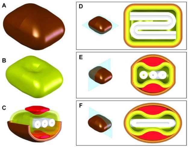
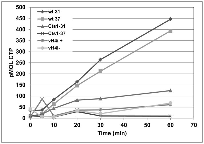
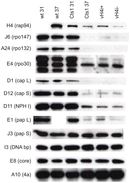
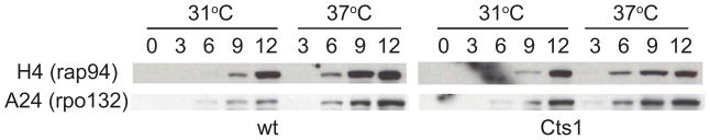
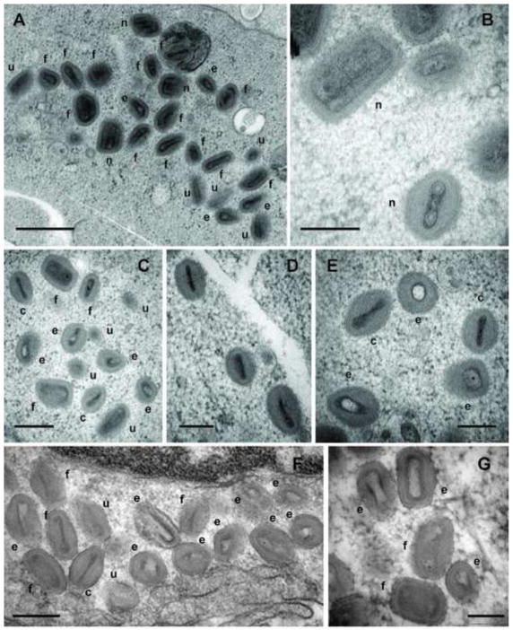
The poxvirus virion contains an inner tubular nucleocapsid structure. The nucleocapsid is apparently labile to conventional electron microscopy fixation procedures and has therefore been largely ignored for decades. Advancements in electron microscopy sample preparation, notably high pressure freezing, better preserve the nucleocapsid structure. Using high pressure freezing and electron microscopy, we have compared the virion structures of wt virus and mutant viruses known to be deficient in packaging of viral transcription enzymes. We show that the mutant viruses lack a defined nucleocapsid. These results support the hypothesis that the nucleocapsid contains the viral DNA genome complexed with viral transcription enzymes and structural proteins. The studies open the door to further investigation of the composition and ultrastructure of the poxvirus nucleocapsid.
Copyright © 2012 Elsevier Inc. All rights reserved.
Figures
Similar articles
-
Vaccinia virus protein A3 is required for the production of normal immature virions and for the encapsidation of the nucleocapsid protein L4.Virology. 2015 Jul;481:1-12. doi: 10.1016/j.virol.2015.02.020. Epub 2015 Mar 9. Virology. 2015. PMID: 25765002 Free PMC article.
-
Vaccinia virus mutations in the L4R gene encoding a virion structural protein produce abnormal mature particles lacking a nucleocapsid.J Virol. 2014 Dec;88(24):14017-29. doi: 10.1128/JVI.02126-14. Epub 2014 Sep 24. J Virol. 2014. PMID: 25253347 Free PMC article.
-
In a nutshell: structure and assembly of the vaccinia virion.Adv Virus Res. 2006;66:31-124. doi: 10.1016/S0065-3527(06)66002-8. Adv Virus Res. 2006. PMID: 16877059 Review.
-
Structural basis for Ebola virus nucleocapsid assembly and function regulated by VP24.Nat Commun. 2025 Mar 10;16(1):2171. doi: 10.1038/s41467-025-57236-4. Nat Commun. 2025. PMID: 40064872 Free PMC article.
-
Vaccinia virus proteolysis--a review.Rev Med Virol. 2006 May-Jun;16(3):187-202. doi: 10.1002/rmv.499. Rev Med Virol. 2006. PMID: 16710840 Free PMC article. Review.
Cited by
-
From crescent to mature virion: vaccinia virus assembly and maturation.Viruses. 2014 Oct 7;6(10):3787-808. doi: 10.3390/v6103787. Viruses. 2014. PMID: 25296112 Free PMC article. Review.
-
Fine structure of the vaccinia virion determined by controlled degradation and immunolocalization.Virology. 2015 Jan 15;475:204-18. doi: 10.1016/j.virol.2014.11.020. Epub 2014 Dec 8. Virology. 2015. PMID: 25486587 Free PMC article.
-
Vaccinia virus protein A3 is required for the production of normal immature virions and for the encapsidation of the nucleocapsid protein L4.Virology. 2015 Jul;481:1-12. doi: 10.1016/j.virol.2015.02.020. Epub 2015 Mar 9. Virology. 2015. PMID: 25765002 Free PMC article.
-
The vaccinia virus E6 protein influences virion protein localization during virus assembly.Virology. 2015 Aug;482:147-56. doi: 10.1016/j.virol.2015.02.056. Epub 2015 Apr 10. Virology. 2015. PMID: 25863879 Free PMC article.
-
Vaccinia virus mutations in the L4R gene encoding a virion structural protein produce abnormal mature particles lacking a nucleocapsid.J Virol. 2014 Dec;88(24):14017-29. doi: 10.1128/JVI.02126-14. Epub 2014 Sep 24. J Virol. 2014. PMID: 25253347 Free PMC article.
References
-
- Ahn BY, Gershon PD, Moss B. RNA polymerase-associated protein Rap94 confers promoter specificity for initiating transcription of vaccinia virus early stage genes. J Biol Chem. 1994;269:7552–7557. - PubMed
-
- Ausubel FM, Brent R, Kingston RE, Moore DD, Seidman JG, Smith JA, Struhl K. Current protocols in molecular biology. John Wiley & Sons; New York: 1994.
-
- Broyles SS. Vaccinia virus transcription. J Gen Virol. 2003;84:2293–2303. - PubMed
Publication types
MeSH terms
Substances
Grants and funding
LinkOut - more resources
Full Text Sources
