

Targeting C/EBP homologous protein with siRNA attenuates cerebral vasospasm after experimental subarachnoid hemorrhage
- PMID: 22944263
- PMCID: PMC3498605
- DOI: 10.1016/j.expneurol.2012.08.025
Targeting C/EBP homologous protein with siRNA attenuates cerebral vasospasm after experimental subarachnoid hemorrhage
Abstract
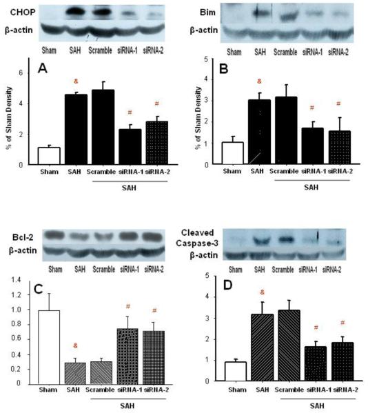
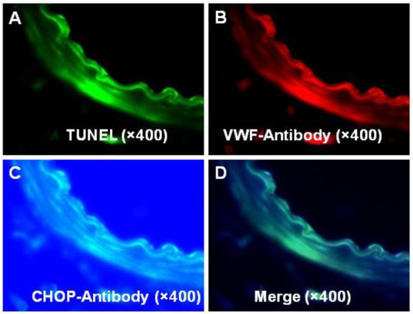
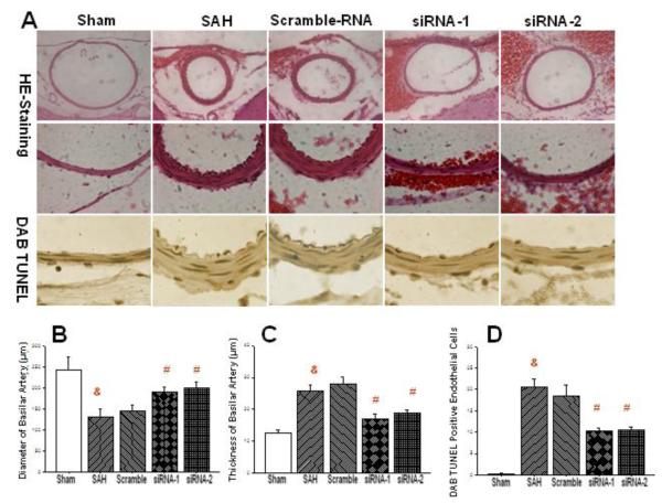
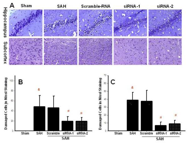
Endothelial apoptosis plays a major role in the development of cerebral vascular spasm after subarachnoid hemorrhage (SAH). C/EBP homologous protein (CHOP) orchestrates apoptosis in a variety of cell types in response to endoplasmic reticulum (ER) stress, implicated in the brain injury after SAH. However, the role of CHOP in the mechanism of cerebral vasospasm (CVS) after SAH remains unexplored. The aim of this study was to evaluate the effect of CHOP silencing on endothelial apoptosis and CVS following subarachnoid hemorrhage in the rat. The study was conducted on 65 rats and employed endovascular perforation model of SAH. CHOP siRNAs were injected 24 h prior to the hemorrhage. At 72 h after SAH brains with basilar arteries (BA) were collected from euthanized rats for laboratory investigations. Triple fluorescence stain revealed expression of CHOP in cerebral vascular endothelia after SAH. Marked reduction of CHOP protein and the reduction of its downstream signaling effectors, bim and caspase-3, were found in BA with Western blot analysis. CHOP silencing reduced number of apoptotic endothelial cells in BA, and increased BA diameter after SAH. The amelioration of CVS was associated with reduced neuronal injury in cerebral tissues. In conclusion, CHOP siRNA treatment can effectively combat apoptotic mechanisms of cerebral vasospasm set in motion by subarachnoid bleeding.
Copyright © 2012 Elsevier Inc. All rights reserved.
Figures
References
-
- Bendel P, Koivisto T, Hanninen T, Kolehmainen A, Kononen M, Hurskainen H, Pennanen C, Vanninen R. Subarachnoid hemorrhage is followed by temporomesial volume loss: MRI volumetric study. Neurology. 2006;67:575–582. - PubMed
-
- Blaschke F, Bruemmer D, Yin F, Takata Y, Wang W, Fishbein MC, Okura T, Higaki J, Graf K, Fleck E, Hsueh WA, Law RE. C-reactive protein induces apoptosis in human coronary vascular smooth muscle cells. Circulation. 2004;110:579–587. - PubMed
-
- Chen G, Zhang S, Shi J, Ai J, Hang C. Effects of recombinant human erythropoietin (rhEPO) on JAK2/STAT3 pathway and endothelial apoptosis in the rabbit basilar artery after subarachnoid hemorrhage. Cytokine. 2009;45:162–168. - PubMed
Publication types
MeSH terms
Substances
Grants and funding
LinkOut - more resources
Full Text Sources
Research Materials
