

Tumor cell vasculogenic mimicry: from controversy to therapeutic promise
- PMID: 22944600
- PMCID: PMC4851740
- DOI: 10.1016/j.ajpath.2012.07.013
Tumor cell vasculogenic mimicry: from controversy to therapeutic promise
Abstract
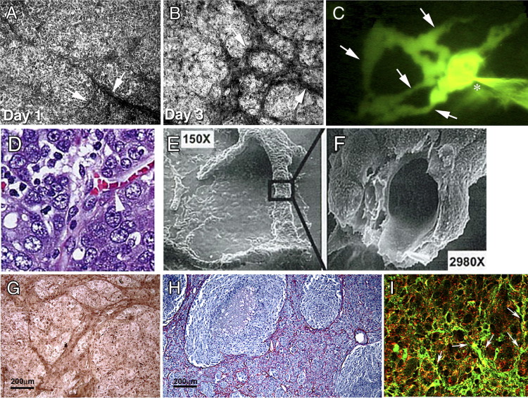
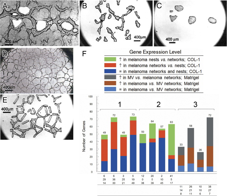
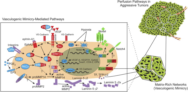
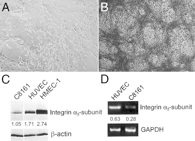
In 1999, The American Journal of Pathology published an article entitled "Vascular channel formation by human melanoma cells in vivo and in vitro: vasculogenic mimicry," by Maniotis and colleagues, which ignited a spirited debate for several years and earned distinction as a citation classic. Tumor cell vasculogenic mimicry (VM) refers to the plasticity of aggressive cancer cells forming de novo vascular networks, which thereby contribute to perfusion of rapidly growing tumors, transporting fluid from leaky vessels, and/or connecting with the constitutional endothelial-lined vasculature. The tumor cells capable of VM share a plastic, transendothelial phenotype, which may be induced by hypoxia. Since VM was introduced as a novel paradigm for melanoma tumor perfusion, many studies have contributed new findings illuminating the underlying molecular pathways supporting VM in a variety of tumors, including carcinomas, sarcomas, glioblastomas, astrocytomas, and melanomas. Facilitating the functional plasticity of tumor cell VM are key proteins associated with vascular, stem cell, and hypoxia-related signaling pathways, each deserving serious consideration as potential therapeutic targets and diagnostic indicators of the aggressive, metastatic phenotype.
Copyright © 2012 American Society for Investigative Pathology. Published by Elsevier Inc. All rights reserved.
Figures
Similar articles
-
Tumor cell vascular mimicry: Novel targeting opportunity in melanoma.Pharmacol Ther. 2016 Mar;159:83-92. doi: 10.1016/j.pharmthera.2016.01.006. Epub 2016 Jan 22. Pharmacol Ther. 2016. PMID: 26808163 Free PMC article. Review.
-
Molecular pathways: vasculogenic mimicry in tumor cells: diagnostic and therapeutic implications.Clin Cancer Res. 2012 May 15;18(10):2726-32. doi: 10.1158/1078-0432.CCR-11-3237. Epub 2012 Apr 2. Clin Cancer Res. 2012. PMID: 22474319 Free PMC article.
-
Mechanisms of vasculogenic mimicry in hypoxic tumor microenvironments.Mol Cancer. 2021 Jan 4;20(1):7. doi: 10.1186/s12943-020-01288-1. Mol Cancer. 2021. PMID: 33397409 Free PMC article. Review.
-
The hypoxia-related signaling pathways of vasculogenic mimicry in tumor treatment.Biomed Pharmacother. 2016 May;80:127-135. doi: 10.1016/j.biopha.2016.03.010. Epub 2016 Mar 21. Biomed Pharmacother. 2016. PMID: 27133049 Review.
-
Vasculogenic mimicry, a complex and devious process favoring tumorigenesis - Interest in making it a therapeutic target.Pharmacol Ther. 2021 Jul;223:107805. doi: 10.1016/j.pharmthera.2021.107805. Epub 2021 Jan 17. Pharmacol Ther. 2021. PMID: 33465401 Review.
Cited by
-
Thymoquinone inhibited vasculogenic capacity and promoted mesenchymal-epithelial transition of human breast cancer stem cells.BMC Complement Med Ther. 2021 Mar 4;21(1):83. doi: 10.1186/s12906-021-03246-w. BMC Complement Med Ther. 2021. PMID: 33663486 Free PMC article.
-
LRIG1 inhibits hypoxia-induced vasculogenic mimicry formation via suppression of the EGFR/PI3K/AKT pathway and epithelial-to-mesenchymal transition in human glioma SHG-44 cells.Cell Stress Chaperones. 2015 Jul;20(4):631-41. doi: 10.1007/s12192-015-0587-y. Epub 2015 Apr 10. Cell Stress Chaperones. 2015. PMID: 25860915 Free PMC article.
-
Matrix metalloproteinases as breast cancer drivers and therapeutic targets.Front Biosci (Landmark Ed). 2015 Jun 1;20(7):1144-63. doi: 10.2741/4364. Front Biosci (Landmark Ed). 2015. PMID: 25961550 Free PMC article. Review.
-
Hinokitiol inhibits vasculogenic mimicry activity of breast cancer stem/progenitor cells through proteasome-mediated degradation of epidermal growth factor receptor.Oncol Lett. 2016 Apr;11(4):2934-2940. doi: 10.3892/ol.2016.4300. Epub 2016 Mar 2. Oncol Lett. 2016. PMID: 27073579 Free PMC article.
-
Tumor cell intravasation.Am J Physiol Cell Physiol. 2016 Jul 1;311(1):C1-C14. doi: 10.1152/ajpcell.00238.2015. Epub 2016 Apr 13. Am J Physiol Cell Physiol. 2016. PMID: 27076614 Free PMC article. Review.
References
Publication types
MeSH terms
Grants and funding
LinkOut - more resources
Full Text Sources
Other Literature Sources
