

MODULATING CO-STIMULATION DURING ANTIGEN PRESENTATION TO ENHANCE CANCER IMMUNOTHERAPY
- PMID: 22945252
- PMCID: PMC3428911
- DOI: 10.2174/187152212802001875
MODULATING CO-STIMULATION DURING ANTIGEN PRESENTATION TO ENHANCE CANCER IMMUNOTHERAPY
Abstract
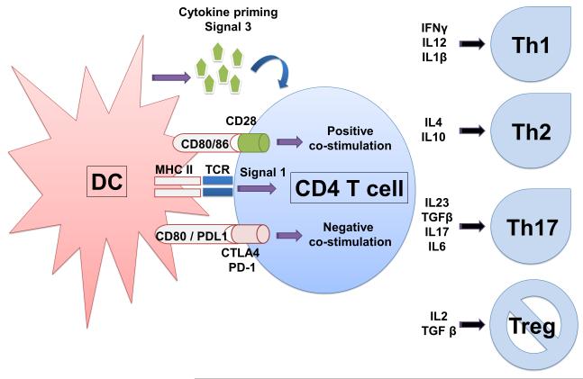
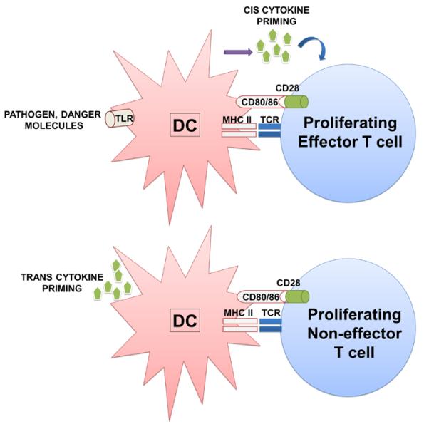
One of the key roles of the immune system is the identification of potentially dangerous pathogens or tumour cells, and raising a wide range of mechanisms to eliminate them from the organism. One of these mechanisms is activation and expansion of antigen-specific cytotoxic T cells, after recognition of antigenic peptides on the surface of antigen presenting cells such as dendritic cells (DCs). However, DCs also process and present autoantigens. Therefore, antigen presentation has to occur in the appropriate context to either trigger immune responses or establishing immunological tolerance. This is achieved by co-stimulation of T cells during antigen presentation. Co-stimulation consists on the simultaneous binding of ligand-receptor molecules at the immunological synapse which will determine the type and extent of T cell responses. In addition, the type of cytokines/chemokines present during antigen presentation will influence the polarisation of T cell responses, whether they lead to tolerance, antibody responses or cytotoxicity. In this review, we will focus on approaches manipulating co-stimulation during antigen presentation, and the role of cytokine stimulation on effective T cell responses. More specifically, we will address the experimental strategies to interfere with negative co-stimulation such as that mediated by PD-L1 (Programmed cell death 1 ligand 1)/PD-1 (Programmed death 1) to enhance anti-tumour immunity.
Figures
References
-
- Morgan RA, Dudley ME, Wunderlich JR, Hughes MS, Yang JC, Sherry RM, Royal RE, Topalian SL, Kammula US, Restifo NP, Zheng Z, Nahvi A, de Vries CR, Rogers-Freezer LJ, Mavroukakis SA, Rosenberg SA. Cancer regression in patients after transfer of genetically engineered lymphocytes. Science. 2006;314(5796):126–129. - PMC - PubMed
-
- Steinman RM, Banchereau J. Taking dendritic cells into medicine. Nature. 2007;449(7161):419–426. - PubMed
-
- Morgado JM, Pratas R, Laranjeira P, Henriques A, Crespo I, Regateiro F, Paiva A. The phenotypical and functional characteristics of cord blood monocytes and CD14(−/low)/CD16(+) dendritic cells can be relevant to the development of cellular immune responses after transplantation. Transpl Immunol. 2008;19(1):55–63. - PubMed
Grants and funding
LinkOut - more resources
Full Text Sources
Other Literature Sources
Research Materials