Review
. 2013 Aug;26(8):987-1003.
doi: 10.1002/nbm.2847. Epub 2012 Sep 4.

A review of calibrated blood oxygenation level-dependent (BOLD) methods for the measurement of task-induced changes in brain oxygen metabolism

Affiliations
Review

A review of calibrated blood oxygenation level-dependent (BOLD) methods for the measurement of task-induced changes in brain oxygen metabolism

Nicholas P Blockley et al. NMR Biomed. 2013 Aug.

Abstract

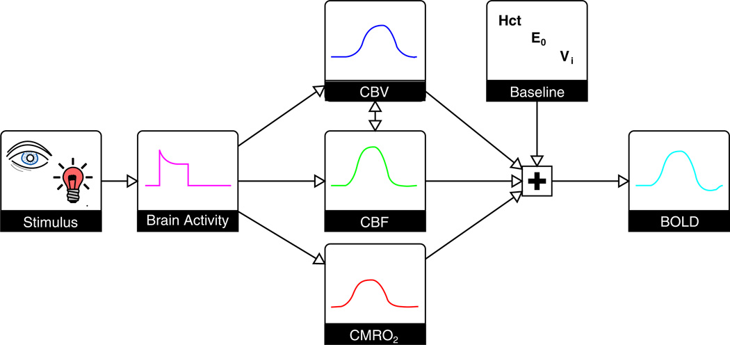
The dynamics of the blood oxygenation level-dependent (BOLD) response are dependent on changes in cerebral blood flow, cerebral blood volume and the cerebral metabolic rate of oxygen consumption. Furthermore, the amplitude of the response is dependent on the baseline physiological state, defined by the haematocrit, oxygen extraction fraction and cerebral blood volume. As a result of this complex dependence, the accurate interpretation of BOLD data and robust intersubject comparisons when the baseline physiology is varied are difficult. The calibrated BOLD technique was developed to address these issues. However, the methodology is complex and its full promise has not yet been realised. In this review, the theoretical underpinnings of calibrated BOLD, and issues regarding this theory that are still to be resolved, are discussed. Important aspects of practical implementation are reviewed and reported applications of this methodology are presented.

Keywords: calibrated BOLD; oxygen metabolism; respiratory challenge; review.


Figures

Figure 1
Figure 1
The blood oxygenation level-dependent (BOLD) response is a complex signal. Basic sensory stimuli elicit an increase in neural activity, resulting in an increase in the cerebral blood flow (CBF) and cerebral metabolic rate of oxygen consumption (CMRO2). CBF increases to a larger degree than CMRO2, and also leads to a local increase in the cerebral blood volume (CBV). The amplitude of the resulting BOLD response is not only dependent on these changes, but also on the baseline physiological state. This baseline is determined by the blood haematocrit (Hct), resting oxygen extraction fraction (OEF) and CBV. An increase in CBF causes an increase in the BOLD signal, whereas increases in venous CBV (CBVv) and CMRO2 cause a decrease. Typically, the CBF effect is dominant, creating a positive BOLD response. The maximum BOLD signal change is determined by the baseline physiological state and increases with increasing Hct, OEF and CBV.
Figure 2
Figure 2
Schematic diagrams of the apparatus typically used to generate a fixed inspired hypercapnia challenge (a) and a fixed inspired hyperoxia challenge (b). In (a), a manually actuated valve enables the inspired gas to be switched between room air and a 5% CO2–air mixture, whereas, in (b), during baseline subjects breathe room air through holes in the mask and hyperoxia is induced by allowing 100% O2 to flow into the mask. Mixing with entrained room air reduces the inspired fraction of oxygen to approximately 50%.
Figure 3
Figure 3
Schematic diagrams of the automated respiratory challenge apparatus currently in use with feedback (a) and feedforward (b) algorithms. The feedback algorithm works by analysing the gas composition of the preceding breath and adjusting the composition of the inflowing fresh gas to force the subject’s end-tidal values towards the targeted values in the following breath. The feedforward algorithm works by calculating the required gas composition to reach the given target end-tidal values using a model of alveolar gas exchange prior to the start of the experiment.
Figure 4
Figure 4
Pulse sequence diagrams for the most common asymmetric spin echo (ASE) methods: (a) single shot ASE; (b) gradient echo sampling of spin echo (GESSE). The methods look very similar, but differ in the way in which phase encoding is applied. For the single shot approach, phase encoding is incremented between k-space traversals in the frequency encode direction using a phase encoding blip. The GESSE method takes a multi-shot approach in which phase encoding is applied prior to the switched frequency encoding gradient. Each lobe of this gradient produces multiple echoes with the same phase encoding. This phase encoding is then incremented across repetitions to fully sample k space.
Figure 5
Figure 5
Simulation of the effect of baseline physiological variability on two calibration methods. (a) Using the standard approach to hyperoxia calibration results in a high degree of uncertainty in the resultant measurement of the cerebral metabolic rate of oxygen consumption (CMRO2) because of the use of an assumed haematocrit (Hct) and resting oxygen extraction fraction (OEF). The blood oxygenation level-dependent (BOLD) signal change normalised by the BOLD scaling parameter (δ S/M) is plotted against the change in cerebral blood flow (CBF) normalised to baseline (CBF0). (b) If these values are known on an individual subject basis, this uncertainty is markedly reduced. (c) For comparison, the standard approach to hypercapnia does not suffer from the same uncertainty as it does not require assumptions about baseline physiology.
Figure 6
Figure 6
Simulation of blood oxygenation level-dependent (BOLD) scaling parameter M for the hypercapnia and R2′ calibration methods. As a result of the effect of diffusion around capillaries, the spin echo refocusing pulse is unable to recover signal lost to dephasing around these vessels. Therefore, R2′ calibration will give a value of M that is lower than that measured using hypercapnia. Further simulations suggest that the required scaling between these methods is relatively stable across different physiological baseline conditions.

References

    1. Ogawa S, Lee TM, Kay AR, Tank DW. Brain magnetic resonance imaging with contrast dependent on blood oxygenation. Proc. Natl. Acad. Sci. USA. 1990;87:9868–9872. - PMC - PubMed
    1. Ogawa S, Tank DW, Menon RS, Ellermann JM, Kim SG, Merkle H, Ugurbil K. Intrinsic signal changes accompanying sensory stimulation: functional brain mapping with magnetic resonance imaging. Proc. Natl. Acad. Sci. USA. 1992;89:5951–5955. - PMC - PubMed
    1. Kwong KK, Belliveau JW, Chesler DA, Goldberg IE, Weisskoff RM, Poncelet BP, Kennedy DN, Hoppel BE, Cohen MS, Turner R. Dynamic magnetic resonance imaging of human brain activity during primary sensory stimulation. Proc. Natl. Acad. Sci. USA. 1992;89:5675–5679. - PMC - PubMed
    1. Bandettini PA, Wong EC, Hinks RS, Tikofsky RS, Hyde JS. Time course EPI of human brain function during task activation. Magn. Reson. Med. 1992;25:390–397. - PubMed
    1. Fox PT, Raichle ME. Focal physiological uncoupling of cerebral blood flow and oxidative metabolism during somatosensory stimulation in human subjects. Proc. Natl. Acad. Sci. USA. 1986;83:1140–1144. - PMC - PubMed

MeSH terms