Review
. 2012 Oct;94(10):841-8.
doi: 10.1002/bdra.23073. Epub 2012 Sep 3.

Does the cranial mesenchyme contribute to neural fold elevation during neurulation?

Affiliations
Review

Does the cranial mesenchyme contribute to neural fold elevation during neurulation?

Irene E Zohn et al. Birth Defects Res A Clin Mol Teratol. 2012 Oct.

Abstract

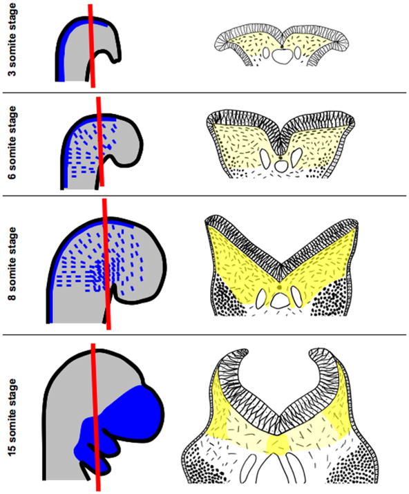
The central nervous system is derived from the neural plate, which undergoes a series of complex morphogenetic events resulting in formation of the neural tube in a process known as neurulation. The cellular behaviors driving neurulation in the cranial region involve forces generated by the neural tissue itself as well as the surrounding epithelium and mesenchyme. Of interest, the cranial mesenchyme underlying the neural plate undergoes stereotypical rearrangements hypothesized to drive elevation of the neural folds. As the neural folds rise, the hyaluronate-rich extracellular matrix greatly expands resulting in increased space between individual cranial mesenchyme cells. Based on inhibitor studies, expansion of the extracellular matrix has been implicated in driving neural fold elevation; however, because the surrounding neural and epidermal ectoderm were also affected by inhibitor exposure, these studies are inconclusive. Similarly, treatment of neurulating embryos with teratogenic doses of retinoic acid results in altered organization of the cranial mesenchyme, but alterations in surrounding tissues are also observed. The strongest evidence for a critical role for the cranial mesenchyme in neural fold elevation comes from studies of genes expressed exclusively in the cranial mesenchyme that when mutated result in exencephaly associated with abnormal organization of the cranial mesenchyme. Twist is the best studied of these and is expressed in both the paraxial mesoderm and neural crest derived cranial mesenchyme. In this article, we review the evidence implicating the cranial mesenchyme in providing a driving force for neural fold elevation to evaluate whether there are sufficient data to support this hypothesis.


Figures

Figure 1
Figure 1. Morphogenesis of the cranial mesenchyme during neural fold elevation
Embryos in whole mount are shown in panels on the left and in sections in panels on the right. The plane of section is illustrated by a red line. In the 3-somite staged mouse embryo, the biconvex neural folds have not yet begun to elevate and exhibit a columnar organization. NC-CM is induced in the dorsal neural tube and can be labeled by the Wnt1-cre driver (blue). Underlying the neural plate are PM-CM cells that can be labeled by the Mesp1-cre line (light grey) and are shown as dashes in panel to the right. Yellow shading in panels to the right indicates the even distribution of HA in the cranial mesenchyme underlying the neural folds. At 6-somite stages, the neuroepithelium transforms to a pseudostratified epithelium and rounded neural crest cells (dots in panel to the right) can be seen migrating from the dorsal neural folds within the subectodermal PM-CM to positions in the branchial arches and frontonasal mesenchyme (blue dashes in left panel). At the 8-somite stage, the neural folds exhibit a “V” shape as they continue to rise and HA concentrations (darker yellow shading) in the ECM increase evenly throughout the cranial mesenchyme. Neural crest cells are migrating and individual PM-CM cells underlying the neural plate are orientated parallel to the neural plate. By the 15-somite stage, NC-CM has migrated to their final destinations in the branchial arches and frontonasal mesenchyme. As the neural folds rise, the HA-rich ECM around the cranial mesenchyme cells in medial regions of the cranial mesenchyme expands resulting in reduced concentration of HA. The neural folds exhibit a “C” shape as the folds begin to converge in the dorsal midline. The PM-CM cells only orientate parallel to the neural tube in the most lateral regions. In medial regions the space between PM-CM cells has expanded greatly. Conceptual diagrams on left were made after Figures 1 and 2 in (Jiang et al., 2002; Yoshida et al., 2008), respectively. Conceptual diagrams on right were made after Figures 2, 4 and 5 in (Morris-Wiman and Brinkley, 1990a).
Figure 2
Figure 2. Failure of neural fold elevation in Hectd1opm mutant embryos is correlated with disorganized cranial mesenchyme
The neural plate in E9.5 Hectd1opm mutant embryos does not transform from the biconvex to “V” then “C” shape demonstrating failure of neural fold elevation. The PM-CM in Hectd1opm mutant embryos fails to undergo the characteristic expansion seen in wildtype embryos and the PM-CM cells underlying the neural plate do not orientate parallel to the neuroepithelium.

Similar articles

Cited by

References

    1. Ackermans MM, et al. Vitamin A and clefting: putative biological mechanisms. Nutr Rev. 2011;69:613–24. - PubMed
    1. Akiyama H, et al. Analytical studies on hyaluronic acid synthesis by normal human epidermal keratinocytes cultured in a serum-free medium. Biol Pharm Bull. 1994;17:361–4. - PubMed
    1. Berk M, et al. Mice lacking the ski proto-oncogene have defects in neurulation, craniofacial, patterning, and skeletal muscle development. Genes Dev. 1997;11:2029–39. - PMC - PubMed
    1. Beverdam A, Meijlink F. Expression patterns of group-I aristaless-related genes during craniofacial and limb development. Mech Dev. 2001;107:163–7. - PubMed
    1. Bildsoe H, et al. Requirement for Twist1 in frontonasal and skull vault development in the mouse embryo. Dev Biol. 2009;331:176–88. - PubMed

Publication types