Review
. 2012 Oct;8(10):610-21.
doi: 10.1038/nrrheum.2012.144. Epub 2012 Sep 4.

The genetics of hyperuricaemia and gout

Affiliations
Review

The genetics of hyperuricaemia and gout

Anthony M Reginato et al. Nat Rev Rheumatol. 2012 Oct.

Abstract

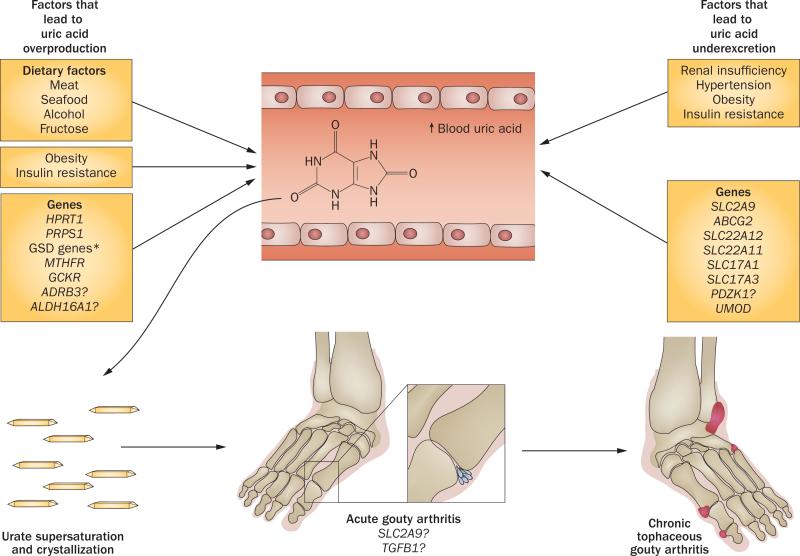
Gout is a common and very painful inflammatory arthritis caused by hyperuricaemia. This review provides an update on the genetics of hyperuricaemia and gout, including findings from genome-wide association studies. Most of the genes that associated with serum uric acid levels or gout are involved in the renal urate-transport system. For example, the urate transporter genes SLC2A9, ABCG2 and SLC22A12 modulate serum uric acid levels and gout risk. The net balance between renal urate absorption and secretion is a major determinant of serum uric acid concentration and loss-of-function mutations in SLC2A9 and SLC22A12 cause hereditary hypouricaemia due to reduced urate absorption and unopposed urate secretion. However, the variance in serum uric acid explained by genetic variants is small and their clinical utility for gout risk prediction seems limited because serum uric acid levels effectively predict gout risk. Urate-associated genes and genetically determined serum uric acid levels were largely unassociated with cardiovascular-metabolic outcomes, challenging the hypothesis of a causal role of serum uric acid in the development of cardiovascular disease. Strong pharmacogenetic associations between HLA-B*5801 alleles and severe allopurinol-hypersensitivity reactions were shown in Asian and European populations. Genetic testing for HLA-B*5801 alleles could be used to predict these potentially fatal adverse effects.


Figures

Figure 1
Figure 1
Genetic variants implicated in the pathogenesis of hyperuricaemia or gout. Discovery timeline showing cumulative number of genes discovered from 2008–2011. Abbreviation: SUA, serum uric acid.
Figure 2
Figure 2
The uric acid transportasome. Urate transporters in renal proximal tubules are involved in the secretion and reabsorption of urate. The balance between these processes determines the net proximal renal excretion. Urate secretion involves SLC22A6 and SLC22A8, which transport uric acid into the epithelial cell across the basolateral membrane, and URAT1, SLC22A13, SLC17A1, SLC17A3, MRP4 and ABCG2, which transport uric acid out of the epithelial cell across the apical membrane. Reabsorption of urate across the apical membrane involves the urate-anion exchangers URAT1 and SLC22A13, which facilitate the entry of urate into the cell in exchange for monocarboxylates (transported into the cell by the sodium-dependent transporters SCL5A8 and SCL5A12) as well as SLC22A11, which exchanges urate and dicarboxylates (transported into the cell by SLC13A3). Antiuricosuric drugs can serve as the exchanging anion for URAT1 and, therefore, enhance urate transport. URAT1 is inhibited by uricosuric agents and might be regulated by hormones. The glucose transporter GLUT-9 also has an important role in reabsorption of urate; GLUT-9b transports uric acid across the apical membrane and GLUT-9a transports uric acid out of the epithelial cell across the basolateral membrane. The scaffolding protein, PDZK1, is involved in the assembly of a transport complex in the apical membrane.
Figure 3
Figure 3
The pathogenesis of hyperuricaemia and gout. Genetic and environmental factors are juxtaposed with the two major mechanisms that lead to hyperuricaemia—exogenous and endogenous overproduction of uric acid and underexcretion of urate. Hyperuricaemia results in the formation of monosodium urate crystals in oversaturated tissue fluids. As well as urate concentration (serum uric acid levels >404.5 μmol/L), crystallization is dependent on pH, temperature and other factors. Stimulation of the NALP3 inflammasome and other humoral and cellular inflammatory mediators by monosodium urate crystals results in acute gouty arthritis. Chronic cumulative urate crystal formation in tissue fluids leads to deposition of monosodium urate crystals in the synovium, cartilage, tendons and soft tissues, resulting in tophi formation and chronic tophaceous gouty arthritis. The vast majority of newly identified common genetic variants that are associated with hyperuricaemia and/or gout are involved in urate renal excretion. However, some of these variants are also expressed in extrarenal tissues and might be involved in the regulation of serum urate homeostasis (ABCG2) or the development of monosodium-urate-crystal-induced inflammation and arthritis (SCL2A9, TGF-β).*Including CPT2, AMP1, ACDS and ALDOB. Abbreviation: GSD, glycogen storage disease.

References

    1. Zhu Y, Pandya BJ, Choi HK. Prevalence of gout and hyperuricaemia in the US general population: The National Health and Nutrition Examination Survey 2007–2008. Arthritis Rheum. 2011;63:3136–3141. - PubMed
    1. Choi HK, Mount DB, Reginato AM, American College of Physicians & American Physiological Society Pathogenesis of gout. Ann. Intern. Med. 2005;143:499–516. - PubMed
    1. Wu EQ, et al. Disease-related and all-cause health care costs of elderly patients with gout. J. Manag. Care Pharm. 2008;14:164–175. - PMC - PubMed
    1. Choi HK, Ford ES, Li C, Curhan G. Prevalence of the metabolic syndrome in patients with gout: the Third National Health and Nutrition Examination Survey. Arthritis Rheum. 2007;57:109–115. - PubMed
    1. Abbott RD, Brand FN, Kannel WB, Castelli WP. Gout and coronary heart disease: the Framingham Study. J. Clin. Epidemiol. 1988;41:237–242. - PubMed

Publication types

MeSH terms