Review
. 2012 Oct;122(10):3476-89.
doi: 10.1172/JCI60777. Epub 2012 Sep 4.

IL-1 receptor antagonist ameliorates inflammasome-dependent alcoholic steatohepatitis in mice

Affiliations
Review

IL-1 receptor antagonist ameliorates inflammasome-dependent alcoholic steatohepatitis in mice

Jan Petrasek et al. J Clin Invest. 2012 Oct.

Abstract

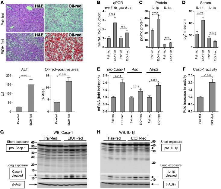
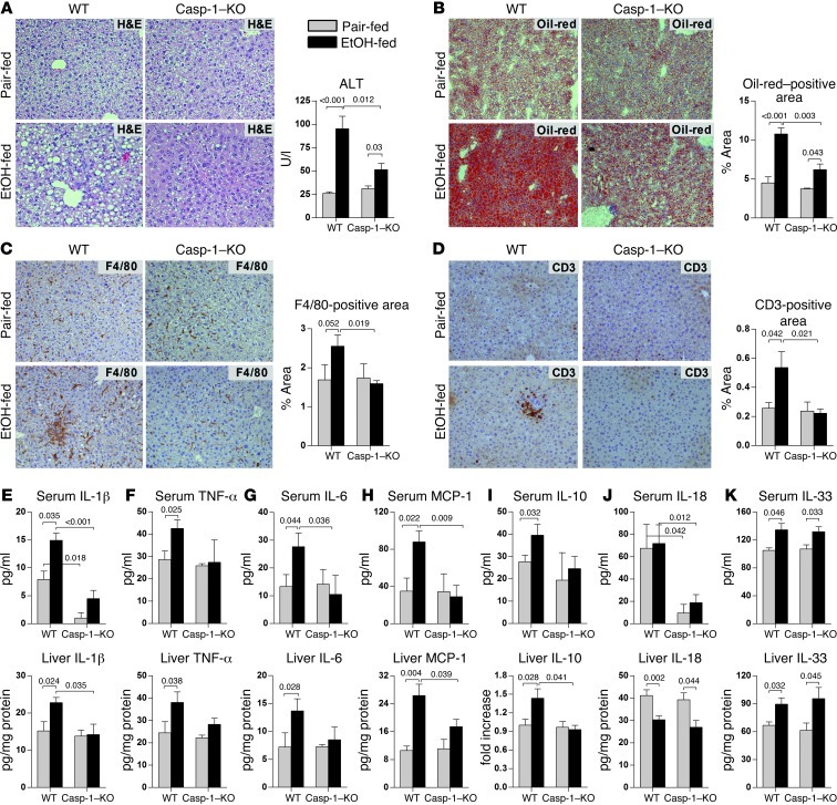
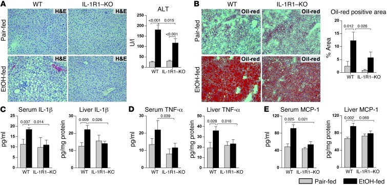
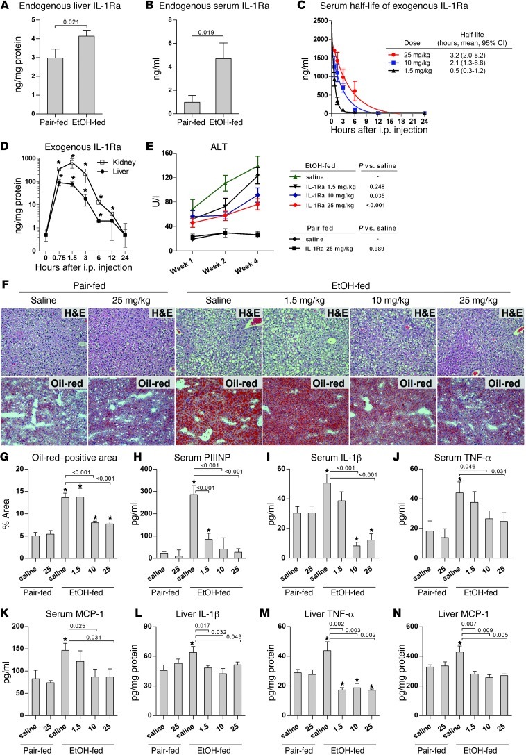
Alcoholic liver disease (ALD) is characterized by steatosis and upregulation of proinflammatory cytokines, including IL-1β. IL-1β, type I IL-1 receptor (IL-1R1), and IL-1 receptor antagonist (IL-1Ra) are all important regulators of the IL-1 signaling complex, which plays a role in inflammation. Furthermore, IL-1β maturation is dependent on caspase-1 (Casp-1). Using IL-1Ra-treated mice as well as 3 mouse models deficient in regulators of IL-1β activation (Casp-1 and ASC) or signaling (IL-1R1), we found that IL-1β signaling is required for the development of alcohol-induced liver steatosis, inflammation, and injury. Increased IL-1β was due to upregulation of Casp-1 activity and inflammasome activation. The pathogenic role of IL-1 signaling in ALD was attributable to the activation of the inflammasome in BM-derived Kupffer cells. Importantly, in vivo intervention with a recombinant IL-1Ra blocked IL-1 signaling and markedly attenuated alcohol-induced liver inflammation, steatosis, and damage. Furthermore, physiological doses of IL-1β induced steatosis, increased the inflammatory and prosteatotic chemokine MCP-1 in hepatocytes, and augmented TLR4-dependent upregulation of inflammatory signaling in macrophages. In conclusion, we demonstrated that Casp-1-dependent upregulation of IL-1β and signaling mediated by IL-1R1 are crucial in ALD pathogenesis. Our findings suggest a potential role of IL-1R1 inhibition in the treatment of ALD.


Figures

Figure 1
Figure 1. Activation of the inflammasome and IL-1β in alcohol-induced liver injury.
WT mice were fed control (pair-fed) or alcohol (EtOH-fed) diet and sacrificed 4 weeks later. Liver samples were stained by H&E or Oil-red-O, and liver injury and steatosis was quantified by measuring serum ALT and Oil-red-O–positive areas, respectively (A). Expression of pro–IL-1β and pro–IL-1α in the liver was analyzed by qPCR (B) and ELISA (C). Secreted forms of IL-1β and IL-1α were measured in the serum using specific ELISA (D). Expression of pro-Casp-1, Asc, and Nlrp3 in the liver was measured using qPCR (E), and Casp-1 activity was measured using a colorimetric assay (F). Cleaved forms of Casp-1 (G) and IL-1β (H) in the livers were analyzed using antibodies that identify both full-length pro-form (short exposure, presented in linear contrast mode) and cleaved forms (long exposure, presented in sigmoidal contrast mode), and normalized to β-actin. See Supplemental Figure 1 for densitometric analysis. n = 5 (pair-fed); 10 (EtOH-fed). Numbers in graphs denote P values. Original magnification, ×200.
Figure 2
Figure 2. Deficiency of Casp-1 attenuates alcoholic liver inflammation, steatosis, and damage.
WT or Casp-1–KO mice were fed control or alcohol diet and sacrificed 4 weeks later. Liver injury was assessed by liver H&E staining and serum ALT (A). Steatosis was evaluated by Oil-red-O staining (B). Immunohistochemistry was used to evaluate recruitment of F4/80-positive macrophages (C) and CD3-positive lymphocytes (D). Serum and liver levels of IL-1β (E), TNF-α (F), IL-6 (G), MCP-1 (H), IL-10 (I), IL-18 (J), and IL-33 (K) were measured as described in Methods. Tissue levels of IL-10 (I; liver) were evaluated using immunoblotting (Supplemental Figure 3A). n = 6 (WT pair-fed); 11 (WT EtOH-fed); 3 (Casp-1–KO pair-fed); 7 (Casp-1–KO EtOH-fed). Numbers in graphs denote P values. Original magnification, ×200.
Figure 3
Figure 3. Deficiency of Casp-1 attenuates alcoholic liver fibrosis.
WT or Casp-1–KO mice were fed control or alcohol diet and sacrificed 4 weeks later. Liver fibrosis was evaluated by Sirius red staining (A) and by measuring expression of Tgfb1 (B) and pro-Col1a1 (C) by qPCR. Specific ELISA was used to evaluate serum markers of liver fibrosis, including PIIINP (D), TIMP-1 (E), and hyaluronic acid (F). n = 6 (WT pair-fed); 11 (WT EtOH-fed); 3 (Casp-1–KO pair-fed); 7 (Casp-1–KO EtOH-fed). Numbers in graphs denote P values. Original magnification, ×200.
Figure 4
Figure 4. Deficiency of ASC attenuates ALD.
WT or ASC-KO mice were fed control or alcohol diet and sacrificed 4 weeks later. Liver injury was assessed by liver H&E staining and serum ALT (A). Steatosis was evaluated by Oil-red-O staining (B). Levels of IL-1β (C), TNF-α (D), IL-6 (E), MCP-1 (F), serum IL-10 (G), IL-18 (H), and IL-33 (I) were measured using specific ELISA; tissue levels of IL-10 (G; liver) were evaluated using immunoblotting (see Supplemental Figure 3B). n = 13 (WT pair-fed); 19 (WT EtOH-fed); 6 (ASC-KO pair-fed); 9 (ASC-KO EtOH-fed). Numbers in graphs denote P values. Original magnification, ×200.
Figure 5
Figure 5. Deficiency in IL-1 signaling in IL-1R1–KOs ameliorates ALD.
WT or IL-1R1–KO mice were fed control (n = 5 per genotype) or alcohol (n = 10 per genotype) diet and sacrificed 4 weeks later. Liver injury was assessed by liver H&E staining and serum ALT (A). Steatosis was evaluated by Oil-red-O staining (B). Serum and liver levels of IL-1β (C), TNF-α (D), and MCP-1 (E) were measured using specific ELISA. Numbers in graphs denote P values. Original magnification, ×200.
Figure 6
Figure 6. Pharmacologic intervention via inhibition of IL-1 signaling ameliorates ALD development.
(A and B) WT mice were fed control (n = 5) or alcohol (n = 10) diet and sacrificed 4 weeks later. Endogenous IL-1Ra was measured in the liver (A) and in the serum (B) using mouse-specific ELISA. (C and D) WT mice (n = 3 per time point and dose) were injected with human recombinant IL-1Ra, and pharmacokinetics was evaluated using human-specific ELISA in the serum (C) and in the liver or kidney extract (D). *P < 0.05 vs. baseline. (EN) WT mice were fed with control (n = 5) or alcohol diet (n = 10 per IL-1Ra dose), treated daily with indicated doses of recombinant human IL-1Ra (anakinra) or saline i.p., and sacrificed 4 weeks later. Liver injury was quantified by serum ALT (E) and H&E (F), and steatosis was evaluated by Oil-red-O staining (F and G). Fibrosis was estimated by PIIINP in the serum (H). Serum and liver levels of IL-1β (I and L), TNF-α (J and M), and MCP-1 (K and N) were measured using specific ELISA. Numbers in graphs denote P values. *P < 0.05 vs. pair-fed saline. Original magnification, ×200.
Figure 7
Figure 7. Pharmacologic intervention via inhibition of IL-1 signaling ameliorates ALD progression.
(AC) WT mice were fed control or alcohol diet. At day 14 (A and B) or day 28 (C), mice were started on daily injections of IL-1Ra (anakinra; 25 mg/kg) or saline i.p. and sacrificed 2 weeks later. Liver histologies from days 14 and 28 were stained with H&E and Oil-red-O (A), and ALT was measured in the serum (B and C). (A and B) n = 5 (pair-fed saline and pair-fed IL-1Ra); 17 (EtOH-fed saline and EtOH-fed IL-1Ra). (C) n = 5 (pair-fed saline and pair-fed IL-1Ra); 9 (EtOH-fed saline and EtOH-fed IL-1Ra). *P < 0.05 vs. pair-fed. (D and E) WT mice were fed control or alcohol diet. After day 14, mice were started on daily injections of IL-1Ra or saline i.p. and sacrificed 5 weeks later. The progression of liver injury was evaluated by measuring serum ALT (D). Liver histologies from days 14 (week 2) and 49 (week 7) were stained with H&E and Oil-red-O (E). n = 5 (pair-fed saline and pair-fed IL-1Ra); 40 (EtOH-fed saline); 29 (EtOH-fed IL-1Ra). At the end of experiment (week 7), survival was 23% in the EtOH-fed saline group and 48% in the EtOH-fed IL-1Ra group (P = 0.036). *P < 0.05 vs. EtOH-fed IL-1Ra; #P<0.05 vs. week 2. Numbers in graphs denote P values. Original magnification, ×200.
Figure 8
Figure 8. Pharmacologic intervention via inhibition of IL-1 signaling facilitates recovery from acute-on-chronic alcoholic liver injury.
WT mice were treated with alcohol diet for 4 weeks and received 3 intragastric gavages of EtOH (5 g/kg) during the last 3 days of alcohol feeding. On day 28, all mice were switched to control diet, and daily treatment with IL-1Ra or saline was initiated. Liver injury was analyzed using serum ALT (A) and H&E staining (B), and steatosis was evaluated by Oil-red-O staining (B). n = 3–5 per time point and treatment. *P < 0.05 vs. baseline. Original magnification, ×200.
Figure 9
Figure 9. BM-derived cells mediate pathogenic effects of Casp-1 in ALD.
(AE) LMNCs or primary hepatocytes were isolated from the livers of chow-fed WT mice as described in Methods. Pro–Casp-1 levels in cell lysate were evaluated using immunoblotting and normalized to β-actin (A). Expression of pro-Casp-1, Asc, Nlrp3, and pro-Il-1b was measured using qPCR (B). WT mice received 1 dose of intragastric EtOH (5 g/kg body weight) or isocaloric dextran-maltose per day on 3 consecutive days. 12 hours after the third intragastric gavage, LMNCs or primary hepatocytes were isolated. Cleaved forms of Casp-1 and IL-1β in cell lysates (C) were analyzed using antibodies that identify both full-length (short exposure, presented in linear contrast mode) and cleaved forms (long exposure, presented in sigmoidal contrast mode) and normalized to β-actin (D and E). LMNCs or hepatocytes were pooled from 5 (A and CE) or 11 (B) mice per group. (FL) WT/WT-BM, Casp-1–KO/WT-BM, and WT/Casp-1–KO–BM mice were fed control (n = 4–5 per genotype) or alcohol (n = 8 per genotype) diet and sacrificed 4 weeks later, as described in Methods. Liver injury was assessed by liver H&E staining and serum ALT (F and H). Steatosis was evaluated by Oil-red-O staining (G and I). Serum levels of IL-1β (J), TNF-α (K), and MCP-1 (L) were measured by specific ELISA. Numbers in graphs denote P values. Original magnification, ×200.
Figure 10
Figure 10. Physiological doses of IL-1β elicit biological response in macrophages and hepatocytes.
(A and B) Immortalized murine RAW264.7 macrophages were stimulated with the indicated doses of recombinant mouse IL-1β (A), or concurrently with recombinant mouse IL-1β and/or LPS (B), and levels of TNF-α were evaluated 12 hours later using specific ELISA. (C and D) Primary hepatocytes isolated from WT mice were treated with recombinant IL-1β. MCP-1 in hepatocyte culture supernatant and in hepatocyte lysate was measured using specific ELISA (C). Triglycerides were measured in primary hepatocytes stimulated for 24 hours with MCP-1 or IL-1β at the indicated doses (D). All stimulations were performed in triplicate. Numbers in graphs denote P values. *P < 0.05 vs. baseline.

Comment in

References

    1. O’Shea RS, Dasarathy S, McCullough AJ. Alcoholic liver disease. Hepatology. 2010;51(1):307–328. doi: 10.1002/hep.23258. - DOI - PubMed
    1. Adachi Y, Bradford BU, Gao W, Bojes HK, Thurman RG. Inactivation of Kupffer cells prevents early alcohol-induced liver injury. Hepatology. 1994;20(2):453–460. doi: 10.1002/hep.1840200227. - DOI - PubMed
    1. Thurman RG. II. Alcoholic liver injury involves activation of Kupffer cells by endotoxin. Am J Physiol. 1998;275(4 pt 1):G605–G611. - PubMed
    1. McClain C, et al. Dysregulated cytokine metabolism, altered hepatic methionine metabolism and proteasome dysfunction in alcoholic liver disease. Alcohol Clin Exp Res. 2005;29(11 suppl):180S–188S. doi: 10.1097/01.alc.0000189276.34230.f5. - DOI - PubMed
    1. Naveau S, et al. A double-blind randomized controlled trial of infliximab associated with prednisolone in acute alcoholic hepatitis. Hepatology. 2004;39(5):1390–1397. doi: 10.1002/hep.20206. - DOI - PubMed

Publication types

MeSH terms