. 2013 Mar;55(1-3):210-6.
doi: 10.1007/s12026-012-8365-8.

TLR7 drives accumulation of ABCs and autoantibody production in autoimmune-prone mice

Affiliations

TLR7 drives accumulation of ABCs and autoantibody production in autoimmune-prone mice

Anatoly V Rubtsov et al. Immunol Res. 2013 Mar.

Abstract

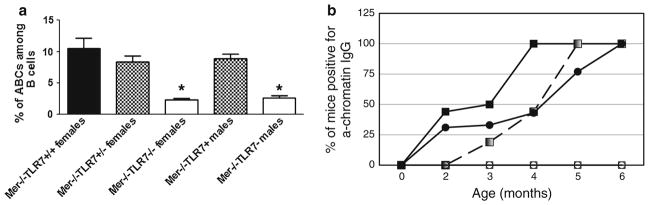
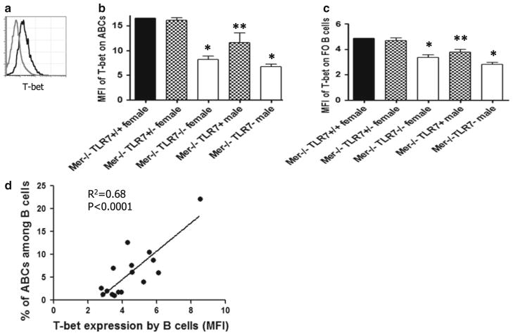
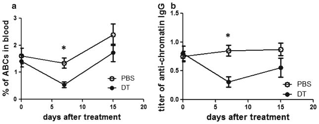
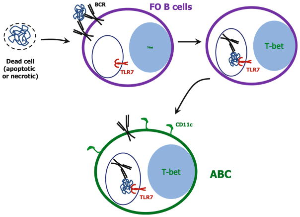
Although autoantibodies are the hallmarks of most autoimmune diseases, the mechanisms by which autoreactive B cells are generated and accumulate are still poorly understood. Overexpression of Toll-like receptor 7 (TLR7) that recognizes single-stranded RNAs has been implicated in systemic lupus erythematosus (SLE), although the cellular mechanism by which this receptor drives the disease is unknown. We recently identified a population of CD11c(+) age-associated B cells (ABCs) which is driven by TLR7 signaling, secretes autoantibodies and appears in autoimmune-prone mice by the time of onset of autoimmunity. Mice lacking the Mer receptor develop autoantibodies and splenomegaly similar to other mouse models of SLE. Here, we show that Mer(-/-) mice that lack TLR7 fail to develop anti-chromatin IgG antibodies, perhaps because they also fail to develop ABCs. Moreover, depletion of CD11c(+) ABCs from Mer(-/-) mice leads to rapid reduction in autoantibodies. Together, these data strongly suggest that ABCs and/or their descendants are the primary source of autoantibodies in Mer(-/-) mice and that TLR7 signaling is crucial for accumulation of ABCs and development of autoantibodies. These data demonstrate for the first time that TLR7, and not TLR9, is responsible for generation of anti-chromatin IgG antibodies in Mer(-/-) mice.


Figures

Fig. 1
Fig. 1
a TLR7 deficiency prevents accumulation of ABCs in Mer−/− mice. Flow cytometry was performed on splenocytes from Mer-deficient mice with various levels of TLR7 expression. At least 5 mice per group were analyzed. ABCs were identified as CD3CD19+B220+CD11c+, and the percentage of ABCs among B cells is demonstrated. Bars represent mean (±SEM) of at least 10 mice per group. *p < 0.001 (Student’s two-tailed t test). b TLR7 deficiency results in the absence of autoantibody accumulation. Serum from Mer−/− TLR7+/+ females (solid squares), Mer−/−TLR7+/− females (gradient squares, dashed line), Mer−/−TLR7−/− females (open squares), Mer−/−TLR7+ males (solid circles), Mer−/−TLR7 males (open circles) at different age was analyzed for the presence of anti-chromatin IgG antibodies, and percentage of mice positive for autoantibodies is shown. The sample was counted positive if the titer was 5 times over the titer of control C57BL/6 mice. At least 10 animals per age group were analyzed
Fig. 2
Fig. 2
a ABCs from Mer-deficient mice express higher level of T-bet than FO B cells. Splenocytes from Mer−/− mice were stained with CD19, B220, CD11c, CD3 and T-bet. ABCs (solid black line) were identified as CD3CD19+B220+CD11c+, and FO B cells (dashed gray line) are CD3CD19+B220+CD11c. Mean fluorescence intensity (MFI) of T-bet expression is shown. Staining is a representative of at least five independent experiments. b T-bet expression in ABCs from Mer−/− mice with different expression of TLR7. Average MFI and standard error is shown for at least 3 mice per group. c T-bet expression in FO B cells from Mer−/− mice. Average MFI and standard error is shown for at least 3 mice per each group. *p < 0.005; **p < 0.05 (Student’s two-tailed t test). d Correlation of percentage of ABCs with the expression of T-bet in all B cells. Linear regression analysis was applied, and statistical significance is demonstrated
Fig. 3
Fig. 3
a Depletion of ABCs in blood by diphtheria toxin in Mer−/− CD11c-DTR/GFP mice. Mice were bled 0, 7 and 15 days after injection with diphtheria toxin or PBS, and blood was analyzed for the presence of ABCs by flow cytometry. Each treatment group contained 5 mice before treatment. b Titer of anti-chromatin IgG autoantibodies in serum of Mer−/− CD11c-DTR/GFP mice was determined 0, 7 and 15 days after injection of diphtheria toxin. Each treatment group contained 5 mice. *p < 0.01 (Student’s two-tailed t test)
Fig. 4
Fig. 4
Model showing nature of appearance of autoantibodies in Mer-deficient mice. Inefficient uptake of dead cells leads to the appearance of autoantigens such as DNA, RNA and nucleoproteins in blood, which in turn is recognized by autoreactive B cells through B cell receptor (BCR). After binding, BCR gets internalized and delivers antigen to endosomes where TLR7 is localized. Recognition of RNA and nucleoproteins delivered by BCR initiates TLR7 signaling leading to the upregulation of T-bet and accumulation of ABCs which are the source of autoantibodies in Mer−/− mice

Similar articles

Cited by

References

    1. Lefkowith JB, Gilkeson GS. Nephritogenic autoantibodies in lupus: current concepts and continuing controversies. Arthr Rheum. 1996;39(6):894–903. - PubMed
    1. Cohen PL, Caricchio R, Abraham V, Camenisch TD, Jennette JC, Roubey RA, et al. Delayed apoptotic cell clearance and lupus-like autoimmunity in mice lacking the c-mer membrane tyrosine kinase. J Exp Med. 2002;196(1):135–40. - PMC - PubMed
    1. Scott RS, McMahon EJ, Pop SM, Reap EA, Caricchio R, Cohen PL, et al. Phagocytosis and clearance of apoptotic cells is mediated by MER. Nature. 2001;411(6834):207–11. - PubMed
    1. Rubtsov AV, Rubtsova K, Kappler JW, Marrack P. Genetic and hormonal factors in female-biased autoimmunity. Autoimmun Rev. 2010;9(7):494–8. - PMC - PubMed
    1. Rubtsov AV, Rubtsova K, Fischer A, Meehan RT, Gillis JZ, Kappler JW, et al. Toll-like receptor 7 (TLR7)-driven accumulation of a novel CD11c B-cell population is important for the development of autoimmunity. Blood. 2011;118(5):1305–15. - PMC - PubMed

Publication types

MeSH terms